. 2021 Nov 10;8(11):210822.
doi: 10.1098/rsos.210822. eCollection 2021 Nov.

Diverse stem-chondrichthyan oral structures and evidence for an independently acquired acanthodid dentition

Affiliations

Diverse stem-chondrichthyan oral structures and evidence for an independently acquired acanthodid dentition

Richard P Dearden et al. R Soc Open Sci. .

Abstract

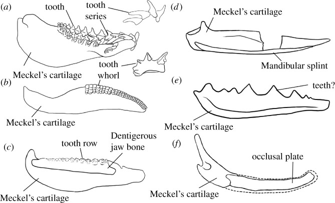
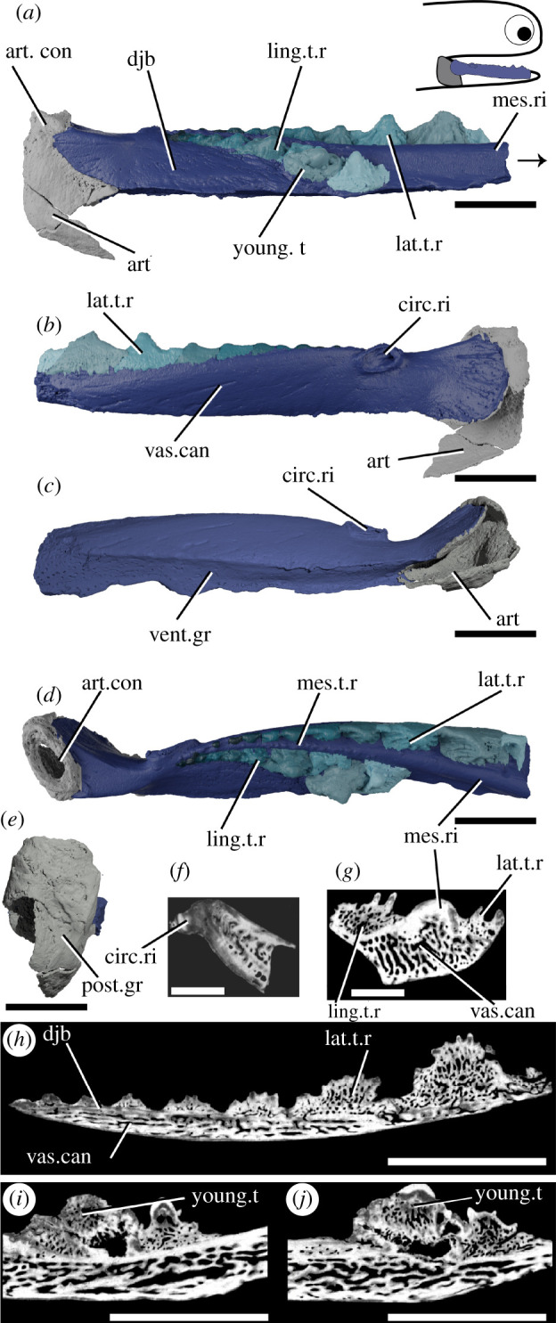
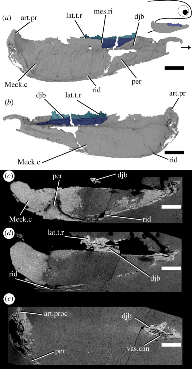
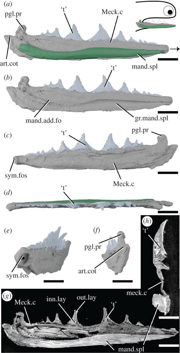
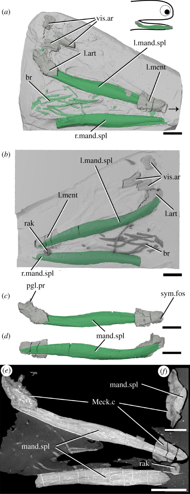
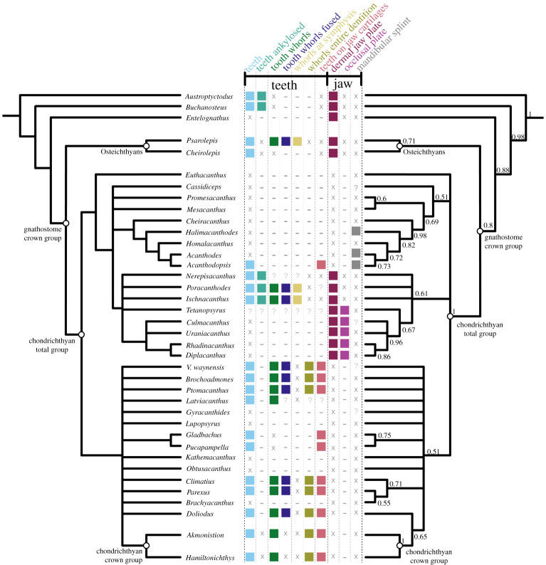
The teeth of sharks famously form a series of transversely organized files with a conveyor-belt replacement that are borne directly on the jaw cartilages, in contrast to the dermal plate-borne dentition of bony fishes that undergoes site-specific replacement. A major obstacle in understanding how this system evolved is the poorly understood relationships of the earliest chondrichthyans and the profusion of morphologically and terminologically diverse bones, cartilages, splints and whorls that they possess. Here, we use tomographic methods to investigate mandibular structures in several early branching 'acanthodian'-grade stem-chondrichthyans. We show that the dentigerous jaw bones of disparate genera of ischnacanthids are united by a common construction, being growing bones with non-shedding dentition. Mandibular splints, which support the ventro-lateral edge of the Meckel's cartilage in some taxa, are formed from dermal bone and may be an acanthodid synapomorphy. We demonstrate that the teeth of Acanthodopsis are borne directly on the mandibular cartilage and that this taxon is deeply nested within an edentulous radiation, representing an unexpected independent origin of teeth. Many or even all of the range of unusual oral structures may be apomorphic, but they should nonetheless be considered when building hypotheses of tooth and jaw evolution, both in chondrichthyans and more broadly.

Keywords: Palaeozoic; acanthodians; chondrichthyans; dentitions; early vertebrates; tooth evolution.


Figures

Figure 1.
Figure 1.
Summary figure showing lower jaws of six chondrichthyans, and major structures discussed in the text. (a) The extant elasmobranch Isurus in medial view and with tooth series in lateral view, drawn from three-dimensional data [31]; (b) the stem-chondrichthyan Ptomacanthus in medial view reconstructed from [32] and with tooth whorl from [21]; (c) the ischnacanthid stem-chondrichthyan Erymnacanthus in medial view redrawn from [25]; (d) the acanthodid stem-chondrichthyan Acanthodes in lateral view redrawn from [33]; (e) the acanthodid stem-chondrichthyan Acanthodopsis in lateral view redrawn from [23]; (f) the diplacanthid stem-chondrichthyan Diplacanthus in medial view reconstructed from [61] and [28]. Not to scale.
Figure 2.
Figure 2.
Tomographic model of the left lower jaw of T. erroli NHMUK PV P.33706 in (a) medial view, (b) lateral view, (c) ventral view, (d) dorsal view, (e) posterior view, and reconstructed tomograms showing (f) a transverse section through the circular ridge, (g) a transverse section through the lower jaw, (h) a sagittal section through the lingual tooth row and (i,j) progressively medial sagittal sections through the aberrant youngest cusp and neighbouring cusps. Teeth in (a), (b) and (d) are coloured separately from the dentigerous jaw bone. Arrow indicates direction of anterior, and top right inset shows location of rendered jaw components. art, articular (Meckel's cartilage); art.con, articular ‘condyle’; circ.ri, circular ridge; djb, dentigerous jaw bone; lat.t.r., lateral tooth row; ling.t.r, lingual tooth row; mes.ri, mesial ridge; mes.t.r, mesial tooth row; post.gr, posterior groove; young.t, out-of-order youngest tooth; vent.gr, ventral groove; vas.can, vascular canals. Scale bar = 5 mm.
Figure 3.
Figure 3.
Tomographic model of a dentigerous jaw bone of Atopacanthus sp. NHMUK PV P.10978 in (a) medial view, (b) lateral view, (c) ventral view, (d) dorsal view and a reconstructed tomograms showing (e) a sagittal section through the lingual tooth row and (f) a transverse section through the lower jaw. Arrow indicates direction of anterior, and top right inset shows possible locations of rendered jaw components. Teeth in (a), (b) and (d) are coloured separately from the dentigerous jaw bone. add.c, additional cusps; djb, dentigerous jaw bone; lat.t.r., lateral tooth row; ling.pl, lingual plate; ling.t.r, lingual tooth row; mes.ri, mesial ridge; mes.t.r, mesial tooth row; vent.gr, ventral groove; vas.can, vascular canals. Scale bar = 5 mm in (ae) and (hj), 2 mm in (fh).
Figure 4.
Figure 4.
Tomographic model of the left lower jaw of Ischnacanthus sp. NHMUK PV P.40124 in (a) lateral view, (b) medial view, and (c), (d), (e) reconstructed tomograms showing successively deeper sagittal sections. Arrow indicates direction of anterior, and top right inset shows location of rendered jaw components. art.proc, articular process; djb, dentigerous jaw bone; lat.t.r., lateral tooth row; Meck.c, Meckel's cartilage; mes.ri, mesial ridge; per, perichondral bone; rid, ridge; vas.can, vascular canal. Scale bar = 5 mm.
Figure 5.
Figure 5.
Tomographic model of the right lower jaw of Acanthodopsis sp. NHMUK PV P.10383 in lateral view with (a) and without (b) mandibular splint, (c) medial view, (d) dorsal view, (e) anteromedial view, (f) posterior view, and reconstructed tomograms showing (g) a sagittal section through the entire jaw and (h) a transverse section through the jaw. Arrow indicates direction of anterior, and top right inset shows location of rendered jaw components. art.cot, articular cotylus; gr.mand.spl, groove for mandibular splint; inn.lay, inner layer; mand.add.fo, mandibular adductor fossa; mand.spl, mandibular splint; Meck.c, Meckel's cartilage; out.lay, outer layer; pgl.pr, preglenoid process; sym.fos, symphyseal fossa; ‘t’, ‘teeth’. Scale bar = 5 mm in (ag), 2 mm in (h).
Figure 6.
Figure 6.
The lower jaws of Acanthodes sp. NHMUK PV P.8065 in (a) dorsal view against the matrix, (b) in ventral view superimposed on a digital mould of the matrix's surface, the left lower jaw isolated in (c) medial and (d) lateral view, and reconstructed tomograms showing (e) a coronal section through the specimen and (f) a transverse section through a lower jaw. Arrow indicates direction of anterior, and top right inset shows location of rendered jaw components. art, articular (Meckel's cartilage); br, branchiostegal rays; mand.spl, mandibular splint; Meck.c, Meckel's cartilage; ment, mentomandibular (Meckel's cartilage); pgl.pr, preglenoid process; rak, gill raker; sym.fos, symphyseal fossa; vis.ar, visceral arch fragments. Scale bar = 5 mm in (a–e), 2 mm in (f).
Figure 7.
Figure 7.
Phylogenetic relationships of early chondrichthyans and distribution of oral structures. Strict consensus of 26,101 most parsimonious trees on left and Bayesian analysis on right, with some non-chondrichthyan taxa excluded (full tree with support values in electronic supplementary material, figures S2 and S3). Numbers at nodes on right represent Bayesian posterior probabilities. Character distribtion is based on coding in the data matrix as follows: teeth, character (c.) 82; teeth ankylosed, c.93; tooth whorls fused, c.85; whorls at symphysis and whorls entire dentition, c.88; teeth on jaw cartilages and dermal jaw plate, c.94; occlusal plate, c.269; mandibular splint, c.268. Filled boxes indicate presence of feature; ‘x’ indicates feature absent; ‘?’ indicates uncertainty; ‘−’ indicates inapplicability.
Figure 8.
Figure 8.
Summaries of four contrasting recent phylogenetic schemes of early chondrichthyans, with distribution of oral characters. (a) King et al. [45], (b) Chevrinais et al. [44], (c) Coates et al. [24], (d) Frey et al. [46]. Icons and character numbers as in figure 7. Acanthodiforms includes taxa both with and without mandibular splints.

Similar articles

Cited by

  • The oldest gnathostome teeth.
    Andreev PS, Sansom IJ, Li Q, Zhao W, Wang J, Wang CC, Peng L, Jia L, Qiao T, Zhu M. Andreev PS, et al. Nature. 2022 Sep;609(7929):964-968. doi: 10.1038/s41586-022-05166-2. Epub 2022 Sep 28. Nature. 2022. PMID: 36171375
  • The skeletal completeness of the Palaeozoic chondrichthyan fossil record.
    Schnetz L, Butler RJ, Coates MI, Sansom IJ. Schnetz L, et al. R Soc Open Sci. 2024 Jan 31;11(1):231451. doi: 10.1098/rsos.231451. eCollection 2024 Jan. R Soc Open Sci. 2024. PMID: 38298400 Free PMC article.

References

    1. Tucker AS, Fraser GJ. 2014. Evolution and developmental diversity of tooth regeneration. Semin. Cell Dev. Biol . 25–26, 71-80. (10.1016/j.semcdb.2013.12.013) - DOI - PubMed
    1. Jobbins M, Rücklin M, Argyriou T, Klug C. 2021. A large middle Devonian eubrachythoracid ‘placoderm’ (Arthrodira) jaw from northern Gondwana. Swiss J. Palaeontol. 140, 2. (10.1186/s13358-020-00212-w) - DOI - PMC - PubMed
    1. Ørvig T. 1980. Histologic studies of ostracoderms, placoderms and fossil elasmobranchs: 3. Structure and growth of the gnathalia of certain arthrodires. Zool. Scr. 9, 141-159. (10.1111/j.1463-6409.1980.tb00660.x) - DOI
    1. Rücklin M, Donoghue PCJ. 2015. Romundina and the evolutionary origin of teeth. Biol. Lett. 11, 20150326. (10.1098/rsbl.2015.0326) - DOI - PMC - PubMed
    1. Rücklin M, Donoghue PCJ, Johanson Z, Trinajstic K, Marone F, Stampanoni M. 2012. Development of teeth and jaws in the earliest jawed vertebrates. Nature 491, 748-751. (10.1038/nature11555) - DOI - PubMed

LinkOut - more resources