

Assessing the Utility of 18F-Fluorodeoxyglucose Positron Emission Tomography in the Differential Diagnosis Between Spinal Schwannomas and Meningiomas
- PMID: 34804734
- PMCID: PMC8599483
- DOI: 10.7759/cureus.18890
Assessing the Utility of 18F-Fluorodeoxyglucose Positron Emission Tomography in the Differential Diagnosis Between Spinal Schwannomas and Meningiomas
Abstract
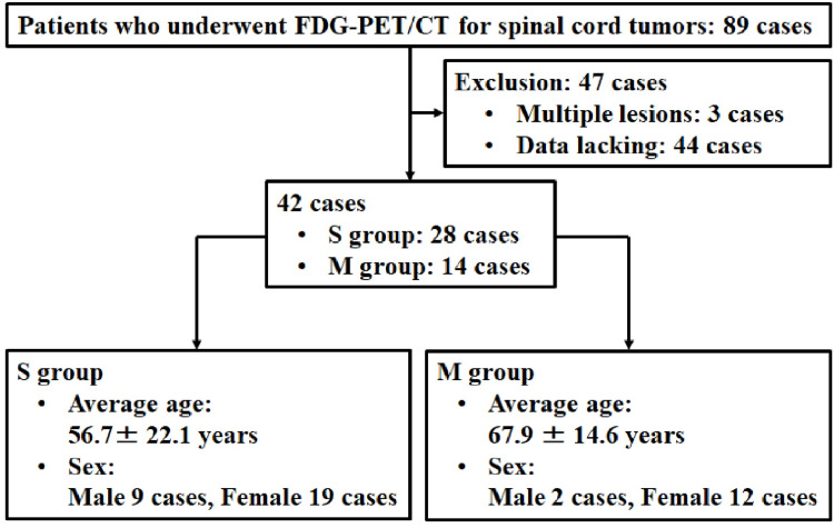
Objective The advantage of 18F-fluorodeoxyglucose-positron emission tomography (FDG-PET) for the differential diagnosis of schwannoma and meningioma remains unclear. The purpose of this study was to compare the maximum standardized uptake value (SUVmax) with computed tomography (CT) and magnetic resonance imaging (MRI) findings and assess its utility in the differential diagnosis of schwannomas and meningiomas. Methods This study included 42 patients who underwent surgery and had pathological diagnoses of schwannomas (S group) or meningiomas (M group). Multivariate logistic regression analyses were conducted using meningioma prevalence as the dependent variable, and confounders were selected from those with p-values <0.05, including calcification, dural tail sign, tumor volume, and SUVmax at each spinal level as independent variables. Results The SUVmax of the spinal canal type at the level of the cervical vertebrae was significantly higher in the M group (4.6 ± 0.8) than in the S group (2.7 ± 1.4; P = 0.017). Multivariate logistic regression analysis showed that the dural tail sign was significantly associated with differential diagnosis between the S and M groups (odds ratio [OR], 0.851; 95% confidence interval [CI], 0.704-1.031, p<0.001). Conclusions The dural tail sign on MRI, but not the SUVmax of FDG-PET, was the most useful for the differential diagnosis between schwannomas and meningiomas.
Keywords: 18f-fluorodeoxyglucose-positron emission tomography; dural tail sign; meningioma; schwannoma; suvmax.
Copyright © 2021, Ono et al.
Conflict of interest statement
The authors have declared that no competing interests exist.
Figures
Similar articles
-
Differentiation between intraspinal schwannoma and meningioma by MR characteristics and clinic features.Radiol Med. 2019 Jun;124(6):510-521. doi: 10.1007/s11547-019-00988-z. Epub 2019 Jan 25. Radiol Med. 2019. PMID: 30684254
-
Prevalence of hitherto unknown brain meningioma detected on 68Ga-DOTATATE positron-emission tomography/computed tomography in patients with metastatic neuroendocrine tumor and exploring potential of 177Lu-DOTATATE peptide receptor radionuclide therapy as single-shot treatment approach targeting both tumors.World J Nucl Med. 2019 Apr-Jun;18(2):160-170. doi: 10.4103/wjnm.WJNM_39_18. World J Nucl Med. 2019. PMID: 31040748 Free PMC article.
-
PET/CT and contrast-enhanced CT imaging findings in benign solitary schwannomas.Eur J Radiol. 2021 Aug;141:109820. doi: 10.1016/j.ejrad.2021.109820. Epub 2021 Jun 9. Eur J Radiol. 2021. PMID: 34139574
-
MR imaging features of spinal schwannomas and meningiomas.J Neuroradiol. 2005 Jan;32(1):42-9. doi: 10.1016/s0150-9861(05)83021-4. J Neuroradiol. 2005. PMID: 15798613
-
18F-fluorodeoxyglucose positron emission tomography (18FDG-PET) for patients with biliary tract cancer: Systematic review and meta-analysis.J Hepatol. 2019 Jul;71(1):115-129. doi: 10.1016/j.jhep.2019.01.038. Epub 2019 Feb 21. J Hepatol. 2019. PMID: 30797051
Cited by
-
Current Knowledge on Spinal Meningiomas Epidemiology, Tumor Characteristics and Non-Surgical Treatment Options: A Systematic Review and Pooled Analysis (Part 1).Cancers (Basel). 2022 Dec 19;14(24):6251. doi: 10.3390/cancers14246251. Cancers (Basel). 2022. PMID: 36551736 Free PMC article. Review.
-
Spinal Meningiomas: A Comprehensive Review and Update on Advancements in Molecular Characterization, Diagnostics, Surgical Approach and Technology, and Alternative Therapies.Cancers (Basel). 2024 Apr 7;16(7):1426. doi: 10.3390/cancers16071426. Cancers (Basel). 2024. PMID: 38611105 Free PMC article. Review.
References
-
- Spinal meningiomas and neurofibromas. BU JW. Acta Radiol. 1953;40:283–300. - PubMed
-
- Intraspinal neurinomas and meningiomas. A clinical survey of 172 cases. Iraci G, Peserico L, Salar G. https://pubmed.ncbi.nlm.nih.gov/5121132/ Int Surg. 1971;56:289–303. - PubMed
-
- Spinal neurofibromas: a report of 66 cases and a comparison with meningiomas. Levy WJ, Latchaw J, Hahn JF, Sawhny B, Bay J, Dohn DF. Neurosurgery. 1986;18:331–334. - PubMed
-
- MR imaging features of spinal schwannomas and meningiomas. De Verdelhan O, Haegelen C, Carsin-Nicol B, et al. J Neuroradiol. 2005;32:42–49. - PubMed
-
- Dural "tail" associated with meningiomas on Gd-DTPA-enhanced MR images: characteristics, differential diagnostic value, and possible implications for treatment. Goldsher D, Litt AW, Pinto RS, Bannon KR, Kricheff II. Radiology. 1990;176:447–450. - PubMed
LinkOut - more resources
Full Text Sources