

Pheochromocytoma and Adrenocortical Carcinoma: Morphological Characteristics in Endoscopic Ultrasound Imaging
- PMID: 34804773
- PMCID: PMC8598390
- DOI: 10.1055/a-1626-1678
Pheochromocytoma and Adrenocortical Carcinoma: Morphological Characteristics in Endoscopic Ultrasound Imaging
Abstract
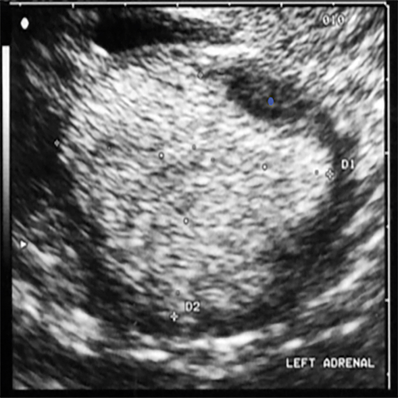
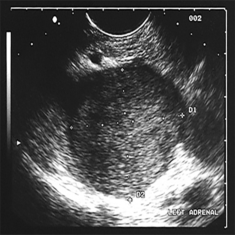
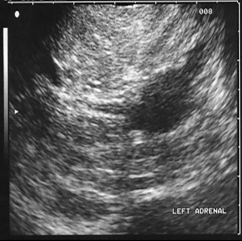
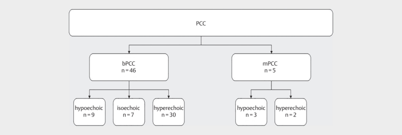
Purpose Pheochromocytoma (PCC) and adrenocortical carcinoma (ACC) are two rare endocrine diseases. Early diagnosis is crucial to significantly reduce morbidity and mortality. In this study, we used endoscopic ultrasound (EUS) for high-resolution imaging to investigate the endosonographic morphology pattern of PCC and ACC. Materials and Methods This retrospective cohort study included 58 PCC/ACC lesions diagnosed by EUS imaging at two tertiary care centers between 1997 and 2015. The following groups were defined by histology or by the presence of a pheochromocytoma-associated syndrome without histological proof: bPCC (benign PCC), mPCC (malignant PCC), and ACC. Results In our cohort, mPCC tended to be larger at the time of diagnosis (n=5; 39.9±41.9 mm) than bPCC (n=46; 27.3 ±20.8 mm, P=0.548). ACC lesions were significantly larger (n=7; 50.6±14.8 mm) than bPCC and mPCC (n=51; 28.5±23.3 mm, P=0.002). In EUS, bPCC and ACC lesions frequently appeared to have a round shape and nodular structure. bPCC and ACC tended to be more hyperechoic (P=0.112 and P=0.558, respectively) and heterogeneous (P=0.501 and P=0.098, respectively) than mPCC. Compared to PCC, ACC did not show high hyperperfusion (P=0.022). In contrast to adenoma, all tumor entities showed hypo-/anechoic areas within the tumor (P<0.05). Conclusion No significant differences in EUS morphology were found to reliably distinguish benign from malignant PCC and ACC lesions. However, EUS may be a reasonable alternative or complementary method to conventional imaging techniques for the early detection of these tumor entities.
Keywords: adrenal gland; tumor; ultrasound.
The Author(s). This is an open access article published by Thieme under the terms of the Creative Commons Attribution-NonDerivative-NonCommercial-License, permitting copying and reproduction so long as the original work is given appropriate credit. Contents may not be used for commercial purposes, or adapted, remixed, transformed or built upon. (https://creativecommons.org/licenses/by-nc-nd/4.0/).
Conflict of interest statement
Conflict of Interest The authors declare that they have no conflict of interest.
Figures
References
-
- Sbardella E, Grossman A B.Pheochromocytoma: An approach to diagnosis. Best Pract Res Clin Endocrinol Metab 2020; 34. doi:10.1016/j.beem.2019.101346 - PubMed
-
- Ahmed A A, Thomas A J, Ganeshan D M.Adrenal cortical carcinoma: pathology, genomics, prognosis, imaging features, and mimics with impact on management. Abdom Radiol 2020; 45. doi:10.1007/s00261-019-02371-y - PubMed
LinkOut - more resources
Full Text Sources