

Ultraviolet photoacoustic microscopy with tissue clearing for high-contrast histological imaging
- PMID: 34804794
- PMCID: PMC8581572
- DOI: 10.1016/j.pacs.2021.100313
Ultraviolet photoacoustic microscopy with tissue clearing for high-contrast histological imaging
Abstract
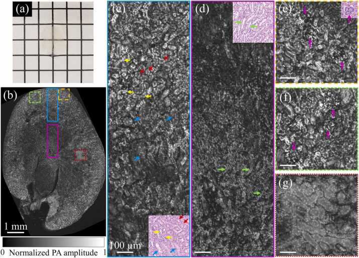
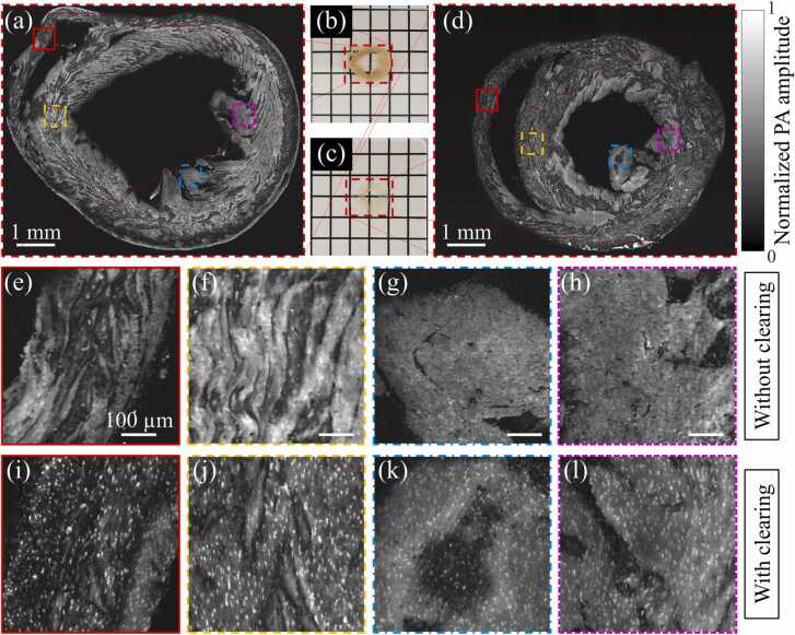
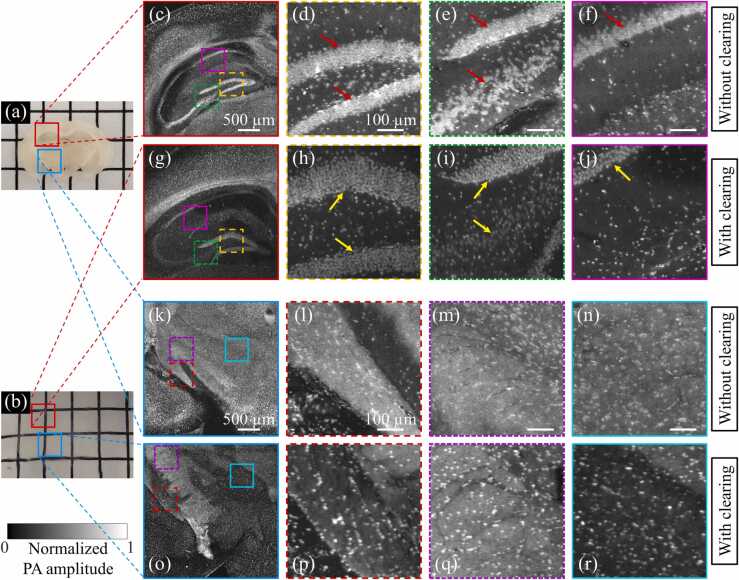
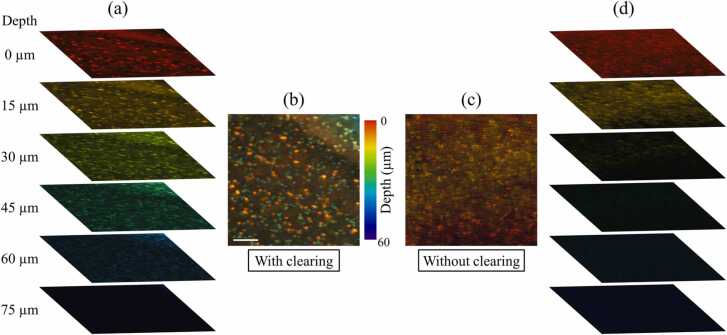
Ultraviolet photoacoustic microscopy (UV-PAM) has been investigated to provide label-free and registration-free volumetric histological images for whole organs, offering new insights into complex biological organs. However, because of the high UV absorption of lipids and pigments in tissue, UV-PAM suffers from low image contrast and shallow image depth, hindering its capability for revealing various microstructures in organs. To improve the UV-PAM imaging contrast and imaging depth, here we propose to implement a state-of-the-art optical clearing technique, CUBIC (clear, unobstructed brain/body imaging cocktails and computational analysis), to wash out the lipids and pigments from tissues. Our results show that the UV-PAM imaging contrast and quality can be significantly improved after tissue clearing. With the cleared tissue, multilayers of cell nuclei can also be extracted from time-resolved PA signals. Tissue clearing-enhanced UV-PAM can provide fine details for organ imaging.
Keywords: Label-free imaging; Photoacoustic microscopy; Tissue clearing; Volumetric imaging.
© 2021 The Authors.
Conflict of interest statement
The authors declare no conflicts of interest.
Figures

Similar articles
-
High-speed label-free ultraviolet photoacoustic microscopy for histology-like imaging of unprocessed biological tissues.Opt Lett. 2020 Oct 1;45(19):5401-5404. doi: 10.1364/OL.401643. Opt Lett. 2020. PMID: 33001904
-
High-throughput ultraviolet photoacoustic microscopy with multifocal excitation.J Biomed Opt. 2018 Mar;23(3):1-6. doi: 10.1117/1.JBO.23.3.036007. J Biomed Opt. 2018. PMID: 29546734 Free PMC article.
-
High-Speed Ultraviolet Photoacoustic Microscopy for Histological Imaging with Virtual-Staining assisted by Deep Learning.J Vis Exp. 2022 Apr 28;(182). doi: 10.3791/63649. J Vis Exp. 2022. PMID: 35575523
-
Volumetric histological characterization of optic nerve degeneration using tissue clearing: literature review and practical study.J Histotechnol. 2021 Dec;44(4):206-216. doi: 10.1080/01478885.2021.1938808. Epub 2021 Jun 16. J Histotechnol. 2021. PMID: 34132156 Review.
-
Photoacoustic microscopy: principles and biomedical applications.Biomed Eng Lett. 2018 Apr 25;8(2):203-213. doi: 10.1007/s13534-018-0067-2. eCollection 2018 May. Biomed Eng Lett. 2018. PMID: 30603203 Free PMC article. Review.
Cited by
-
Review of low-cost light sources and miniaturized designs in photoacoustic microscopy.J Biomed Opt. 2024 Jan;29(Suppl 1):S11503. doi: 10.1117/1.JBO.29.S1.S11503. Epub 2023 Oct 20. J Biomed Opt. 2024. PMID: 37869479 Free PMC article. Review.
-
Dual-wavelength UV-visible metalens for multispectral photoacoustic microscopy: A simulation study.Photoacoustics. 2023 Aug 16;32:100545. doi: 10.1016/j.pacs.2023.100545. eCollection 2023 Aug. Photoacoustics. 2023. PMID: 37645253 Free PMC article.
-
Functional photoacoustic microscopy of hemodynamics: a review.Biomed Eng Lett. 2022 Apr 10;12(2):97-124. doi: 10.1007/s13534-022-00220-4. eCollection 2022 May. Biomed Eng Lett. 2022. PMID: 35529339 Free PMC article. Review.
-
Recent advancements in molecular photoacoustic tomography.JPhys Photonics. 2025 Jul 31;7(3):032003. doi: 10.1088/2515-7647/adf167. Epub 2025 Jul 28. JPhys Photonics. 2025. PMID: 40734710 Free PMC article. Review.
-
A comprehensive review of high-performance photoacoustic microscopy systems.Photoacoustics. 2025 Jun 4;44:100739. doi: 10.1016/j.pacs.2025.100739. eCollection 2025 Aug. Photoacoustics. 2025. PMID: 40528993 Free PMC article. Review.
References
-
- Murray E., Cho J.H., Goodwin D., Ku T., Swaney J., Kim S.Y., Choi H., Park Y.G., Park J.Y., Hubbert A., McCue M., Vassallo S., Bakh N., Frosch M.P., Wedeen V.J., Seung H.S., Chung K. Simple, scalable proteomic imaging for high-dimensional profiling of intact systems. Cell. 2015;163(6):1500–1514. - PMC - PubMed
-
- Chung K., Wallace J., Kim S.Y., Kalyanasundaram S., Andalman A.S., Davidson T.J., Mirzabekov J.J., Zalocusky K.A., Mattis J., Denisin A.K., Pak S., Bernstein H., Ramakrishnan C., Grosenick L., Gradinaru V., Deisseroth K. Structural and molecular interrogation of intact biological systems. Nature. 2013;497(7449):332–337. - PMC - PubMed
-
- Kubota S.I., Takahashi K., Nishida J., Morishita Y., Ehata S., Tainaka K., Miyazono K., Ueda H.R. Whole-body profiling of cancer metastasis with single-cell resolution. Cell Rep. 2017;20(1):236–250. - PubMed
-
- Tanaka N., Kanatani S., Tomer R., Sahlgren C., Kronqvist P., Kaczynska D., Louhivuori L., Kis L., Lindh C., Mitura P., Stepulak A., Corvigno S., Hartman J., Micke P., Mezheyeuski A., Strell C., Carlson J.W., Fernández Moro C., Dahlstrand H., Östman A., Matsumoto K., Wiklund P., Oya M., Miyakawa A., Deisseroth K., Uhlén P. Whole-tissue biopsy phenotyping of three-dimensional tumours reveals patterns of cancer heterogeneity. Nat. Biomed. Eng. 2017;1(10):796–806. - PubMed
LinkOut - more resources
Full Text Sources
Miscellaneous
