

Primary Giant Cell Tumor of the Breast With Pulmonary Metastasis: A Case Report and Review of the Literature
- PMID: 34804910
- PMCID: PMC8602786
- DOI: 10.3389/fonc.2021.638237
Primary Giant Cell Tumor of the Breast With Pulmonary Metastasis: A Case Report and Review of the Literature
Abstract
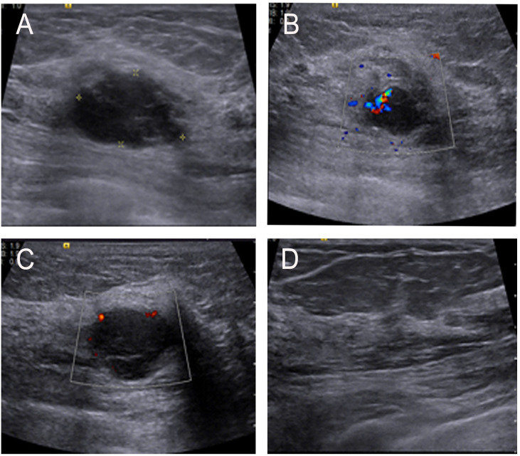
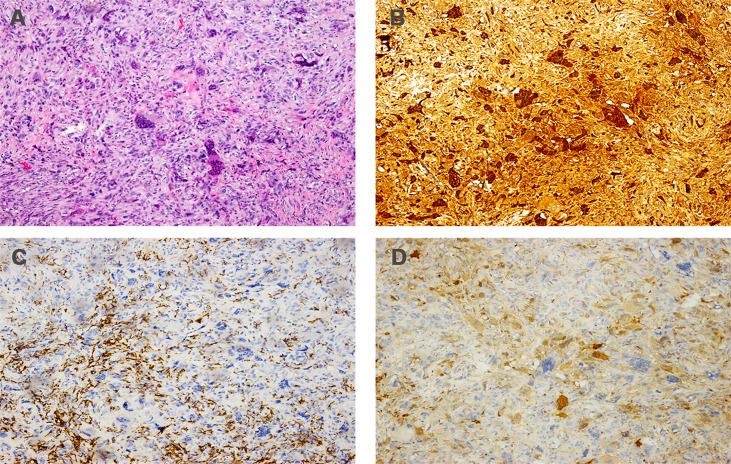
Giant cell tumor of soft tissue (GCT-ST) is an extremely rare tumor that is similar in morphology and immunohistochemistry to giant cell tumor of the bone. Almost 80% of these tumors occur in the upper and lower extremities, and the breast is a very rare location. Here, we report a case of a 65-year-old female patient with a small mobile palpable lump in the left breast. Although the left breast tumor was considered malignant on preoperative imaging, no evidence of malignant tumor was found by ultrasound-guided core needle biopsy (CNB). Subsequently, the left breast tumor was confirmed as a malignant tumor by intraoperative rapid pathological examination. The initial treatment of the tumor was wide local excision and sentinel lymph node biopsy, and it was confirmed to be GCT-ST by histopathology and immunohistochemistry. Despite surgical treatment achieving clear surgical margins, the patient experienced lung metastases within a year of her initial treatment. Fortunately, the patient underwent surgical treatment of lung metastases, and at the last follow-up, the patient was still alive. This is the first case of a primary soft tissue tumor of the breast that has undergone surgical intervention after lung metastasis. This case report highlights the complexity of the clinical diagnosis and treatment of GCT-ST arising from the breast. Surgery may be another good treatment when the patient develops lung metastases.
Keywords: breast tumor; diagnosis; giant cell tumor of soft tissue; prognosis; treatment.
Copyright © 2021 Zhang, Kong, Qi, Wang, Liu, Fang, Song and Wang.
Conflict of interest statement
The authors declare that the research was conducted in the absence of any commercial or financial relationships that could be construed as a potential conflict of interest.
Figures


Similar articles
-
Multimodal Ultrasound Imaging in the Diagnosis of Primary Giant Cell Tumor of the Breast: A Case Report and Literature Review.J Clin Ultrasound. 2025 May;53(4):885-892. doi: 10.1002/jcu.23902. Epub 2024 Dec 9. J Clin Ultrasound. 2025. PMID: 39653506 Review.
-
Preoperative diagnosis of a giant cell tumor of soft tissue arising from the breast by ultrasound-guided core needle biopsy.J Med Ultrason (2001). 2019 Apr;46(2):257-261. doi: 10.1007/s10396-018-0891-0. Epub 2018 Jul 30. J Med Ultrason (2001). 2019. PMID: 30062496
-
A case of giant cell tumor of the breast, clinically suspected as malignant breast tumor.Surg Case Rep. 2019 May 10;5(1):77. doi: 10.1186/s40792-019-0635-4. Surg Case Rep. 2019. PMID: 31076887 Free PMC article.
-
Giant cell tumor of soft tissue arising in breast.Ann Diagn Pathol. 2007 Oct;11(5):345-9. doi: 10.1016/j.anndiagpath.2006.03.013. Epub 2007 Jul 24. Ann Diagn Pathol. 2007. PMID: 17870021
-
Recurrent primary mediastinal giant cell tumor of soft tissue with radiological findings: a rare case report and literature review.World J Surg Oncol. 2017 Jul 26;15(1):137. doi: 10.1186/s12957-017-1205-5. World J Surg Oncol. 2017. PMID: 28747182 Free PMC article. Review.
References
Publication types
LinkOut - more resources
Full Text Sources