

Perifosine, a Bioavailable Alkylphospholipid Akt Inhibitor, Exhibits Antitumor Activity in Murine Models of Cancer Brain Metastasis Through Favorable Tumor Exposure
- PMID: 34804943
- PMCID: PMC8600181
- DOI: 10.3389/fonc.2021.754365
Perifosine, a Bioavailable Alkylphospholipid Akt Inhibitor, Exhibits Antitumor Activity in Murine Models of Cancer Brain Metastasis Through Favorable Tumor Exposure
Abstract
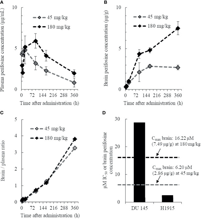
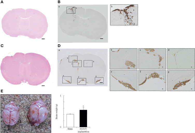
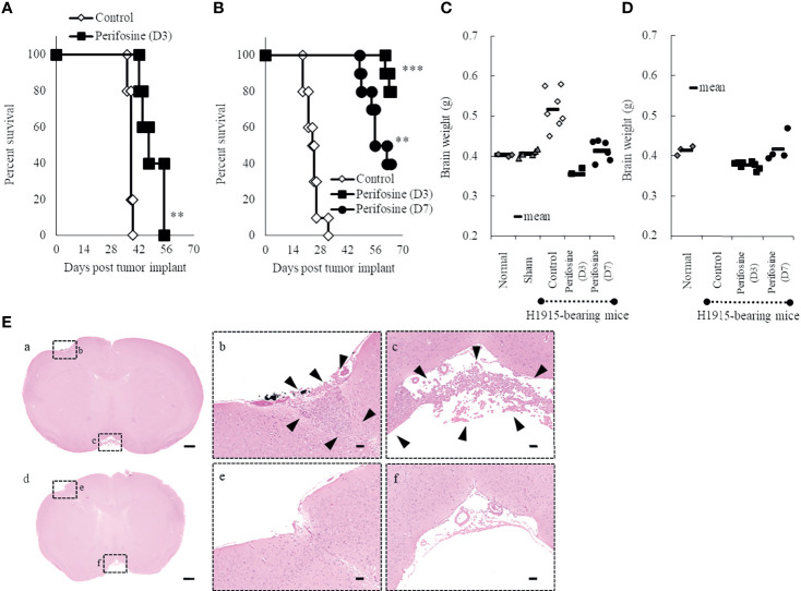
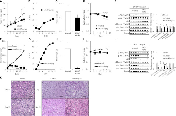
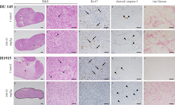
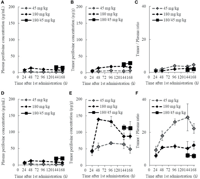
Metastatic brain tumors are regarded as the most advanced stage of certain types of cancer; however, chemotherapy has played a limited role in the treatment of brain metastases. Here, we established murine models of brain metastasis using cell lines derived from human brain metastatic tumors, and aimed to explore the antitumor efficacy of perifosine, an orally active allosteric Akt inhibitor. We evaluated the effectiveness of perifosine by using it as a single agent in ectopic and orthotopic models created by injecting the DU 145 and NCI-H1915 cell lines into mice. Initially, the injected cells formed distant multifocal lesions in the brains of NCI-H1915 mice, making surgical resection impractical in clinical settings. We determined that perifosine could distribute into the brain and remain localized in that region for a long period. Perifosine significantly prolonged the survival of DU 145 and NCI-H1915 orthotopic brain tumor mice; additionally, complete tumor regression was observed in the NCI-H1915 model. Perifosine also elicited much stronger antitumor responses against subcutaneous NCI-H1915 growth; a similar trend of sensitivity to perifosine was also observed in the orthotopic models. Moreover, the degree of suppression of NCI-H1915 tumor growth was associated with long-term exposure to a high level of perifosine at the tumor site and the resultant blockage of the PI3K/Akt signaling pathway, a decrease in tumor cell proliferation, and increased apoptosis. The results presented here provide a promising approach for the future treatment of patients with metastatic brain cancers and emphasize the importance of enriching a patient population that has a higher probability of responding to perifosine.
Keywords: AKT; PI3K; apoptosis; brain cancer; metastasis; signaling pathway.
Copyright © 2021 Taniguchi, Suzuki, Okamura, Kurita, Nohara, Ishii, Kado, Takagi, Tsugane and Shishido.
Conflict of interest statement
KT, TS, TO, AK, GN, SI, SK, AT, MT, and YS are employed by Yakult Honsha Co., Ltd.
Figures
Similar articles
-
The alkylphospholipid perifosine induces apoptosis of human lung cancer cells requiring inhibition of Akt and activation of the extrinsic apoptotic pathway.Mol Cancer Ther. 2007 Jul;6(7):2029-38. doi: 10.1158/1535-7163.MCT-07-0004. Epub 2007 Jun 29. Mol Cancer Ther. 2007. PMID: 17604333
-
Preclinical evaluation of perifosine as a potential promising anti-rhabdomyosarcoma agent.Tumour Biol. 2016 Jan;37(1):1025-33. doi: 10.1007/s13277-015-3740-4. Epub 2015 Aug 13. Tumour Biol. 2016. PMID: 26269112
-
Proapoptotic activity and chemosensitizing effect of the novel Akt inhibitor perifosine in acute myelogenous leukemia cells.Leukemia. 2008 Jan;22(1):147-60. doi: 10.1038/sj.leu.2404980. Epub 2007 Oct 11. Leukemia. 2008. PMID: 17928881
-
Enhancing perifosine's anticancer efficacy by preventing autophagy.Autophagy. 2010 Jan;6(1):184-5. doi: 10.4161/auto.6.1.10816. Epub 2010 Jan 1. Autophagy. 2010. PMID: 20023422 Review.
-
Akt Pathway Inhibitors.Curr Top Med Chem. 2020;20(10):883-900. doi: 10.2174/1568026620666200224101808. Curr Top Med Chem. 2020. PMID: 32091335 Review.
Cited by
-
ISOC1 Modulates Inflammatory Responses in Macrophages through the AKT1/PEX11B/Peroxisome Pathway.Molecules. 2022 Sep 11;27(18):5896. doi: 10.3390/molecules27185896. Molecules. 2022. PMID: 36144632 Free PMC article.
-
Corneal ring infiltrate- far more than Acanthamoeba keratitis: review of pathophysiology, morphology, differential diagnosis and management.J Ophthalmic Inflamm Infect. 2023 Dec 19;13(1):55. doi: 10.1186/s12348-023-00379-6. J Ophthalmic Inflamm Infect. 2023. PMID: 38112842 Free PMC article. Review.
-
The combination of temozolomide and perifosine synergistically inhibit glioblastoma by impeding DNA repair and inducing apoptosis.Cell Death Discov. 2024 Jul 8;10(1):315. doi: 10.1038/s41420-024-02085-1. Cell Death Discov. 2024. PMID: 38977680 Free PMC article.
References
-
- Navarro-Olvera JL, Ariñez-Barahona E, Esqueda-Liquidano MA, Muñoz-Cobos A. Brain Metastases: Literature Review. Rev Med Hosp Gen Méx (2017) 80:60–6. doi: 10.1016/j.hgmx.2016.04.006 - DOI
-
- Nakayama A, Takagi S, Yusa T, Yaguchi M, Hayashi A, Tamura T, et al. . Antitumor Activity of TAK-285, an Investigational, Non-Pgp Substrate HER2/EGFR Kinase Inhibitor, in Cultured Tumor Cells, Mouse and Rat Xenograft Tumors, and in an HER2-Positive Brain Metastasis Model. J Cancer (2013) 4:557–65. doi: 10.7150/jca.6689 - DOI - PMC - PubMed
LinkOut - more resources
Full Text Sources