

Pan-Cancer Analysis of IGF-1 and IGF-1R as Potential Prognostic Biomarkers and Immunotherapy Targets
- PMID: 34804946
- PMCID: PMC8602838
- DOI: 10.3389/fonc.2021.755341
Pan-Cancer Analysis of IGF-1 and IGF-1R as Potential Prognostic Biomarkers and Immunotherapy Targets
Abstract
Aim: Insulin-like growth factor-1 receptor (IGF-1R) is one of the main members of the tyrosine protein kinase receptor family. This receptor binds insulin-like growth factor-1 (IGF-1) with a high affinity. IGF-1 is a member of a family of proteins involved in mediating growth and development. However, the correlations of IGF-1 and IGF-1R to prognosis and tumor-infiltrating lymphocytes in different cancers remain unclear.
Method: This research comprehensively analyzed the expression pattern of IGF-1 and IGF-1R and the influence of IGF-1 and IGF-1R on clinical significance in prognosis prediction among 33 types of malignancies using The Cancer Genome Atlas (TCGA) and the Cancer Cell Line Encyclopedia (CCLE) databases. The correlation between IGF-1, IGF-1R, and cancer immunity was explored.
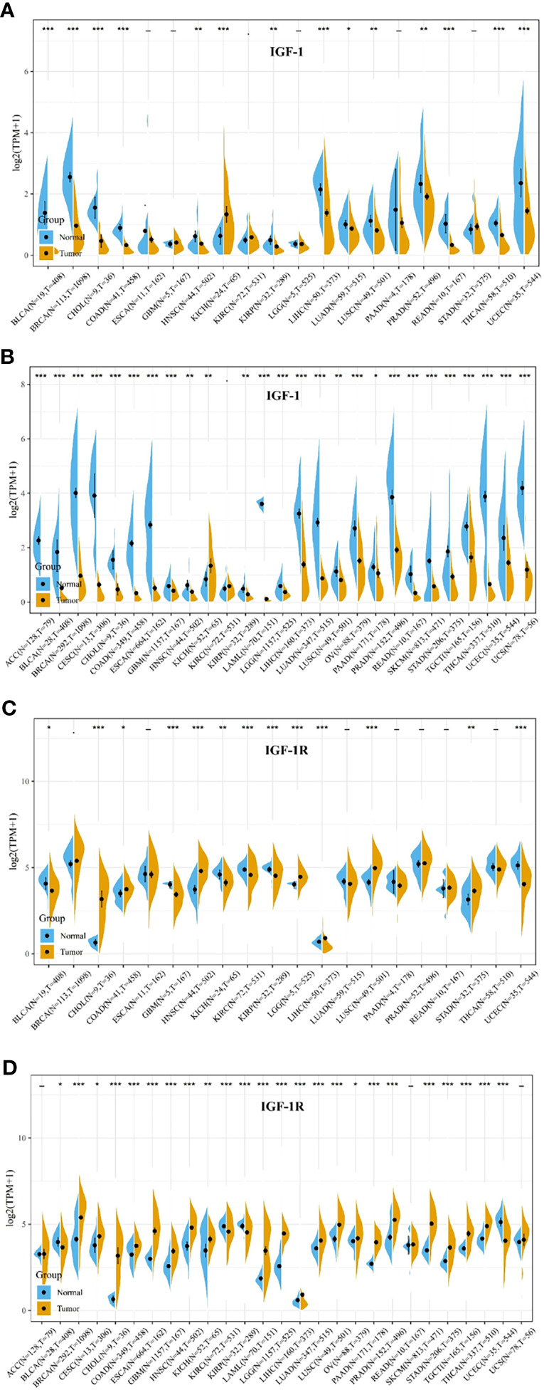
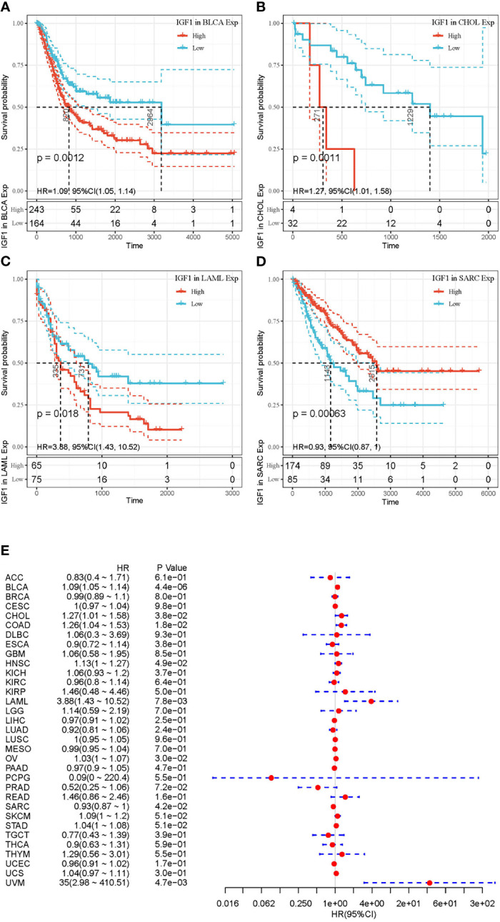
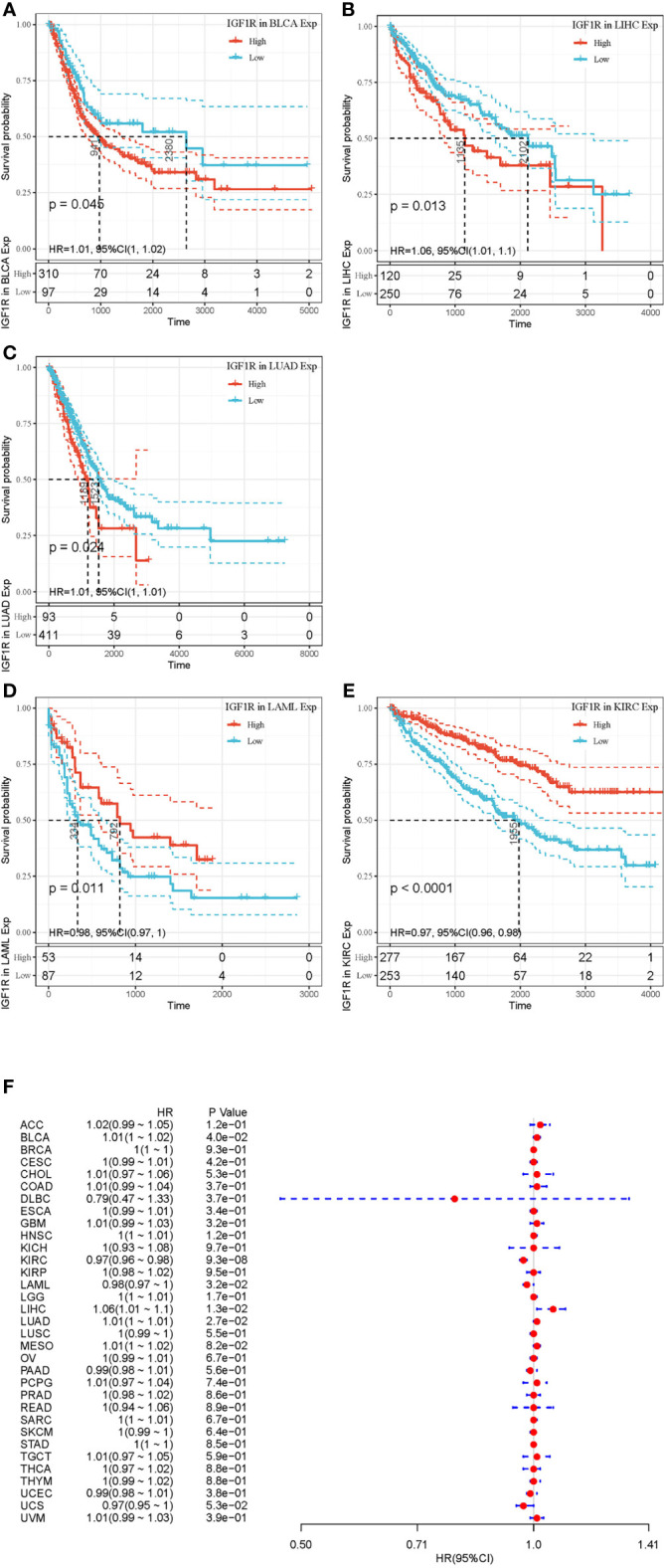
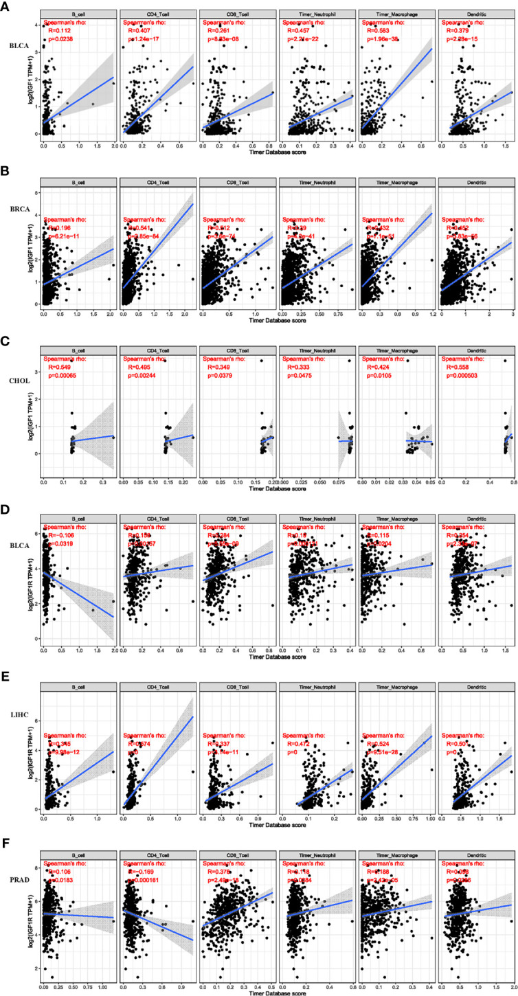
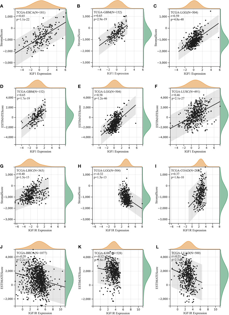
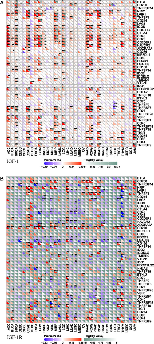
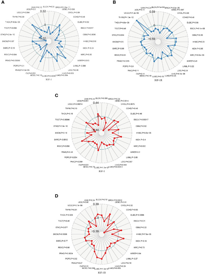
Results: IGF-1 and IGF-1R displayed inconsistent gene expression levels among diverse cancer cell lines. Typically, high expression level of IGF-1 and IGF-1R was detected in most malignant tumors. High expression of IGF-1 was closely bound up with the unfavorable overall survival (OS) for patients in BLCA, CHOL, and LAML upon Cox and Kaplan-Meier analyses. While high expression of IGF-1R was closely bound up with the unfavorable overall survival (OS) for patients in BLCA, LIHC, and LUAD. Furthermore, high expression level of IGF-1 and IGF-1R were closely connected with high degrees of tumor infiltrates, including CD4+ T cell, dendritic cells, and macrophages. In addition, we found that IGF-1 was commonly positively correlated with the expression of gene markers including LAIR1, ICOS, CD40LG, CTLA4, CD48, CD28, CD200R1, HAVCR2, and CD86. Whereas, IGF-1R was commonly positively correlated with the expression of gene markers including NRP1 and CD276. More importantly, IGF-1 and IGF-1R expression were correlated with tumor mutation burden (TMB), microsatellite instability (MSI), mismatch repair (MMR), and DNA methyltransferase (DNMT) of different types of cancers.
Conclusions: The impact of high IGF-1 and IGF-1R on prognosis and immune infiltrates differs across cancer types. Anti-IGF-1R therapy may inhibit tumor growth and contribute to immunotherapy in LIHC and KIRC.
Keywords: IGF-1; IGF-1R; immunity; pan-cancer; prognostic biomarker.
Copyright © 2021 Zhang, Gao, Cao, Wu, Chen, Han, Mo, Qiu, Fan, Zhou and Shen.
Conflict of interest statement
The authors declare that the research was conducted in the absence of any commercial or financial relationships that could be construed as a potential conflict of interest.
Figures
Similar articles
-
Pan-Cancer Analysis of PARP1 Alterations as Biomarkers in the Prediction of Immunotherapeutic Effects and the Association of Its Expression Levels and Immunotherapy Signatures.Front Immunol. 2021 Aug 31;12:721030. doi: 10.3389/fimmu.2021.721030. eCollection 2021. Front Immunol. 2021. PMID: 34531868 Free PMC article.
-
Pan-Cancer Analysis Based on EPOR Expression With Potential Value in Prognosis and Tumor Immunity in 33 Tumors.Front Oncol. 2022 Mar 14;12:844794. doi: 10.3389/fonc.2022.844794. eCollection 2022. Front Oncol. 2022. PMID: 35359375 Free PMC article.
-
Pan-Cancer Analysis Revealed SRSF9 as a New Biomarker for Prognosis and Immunotherapy.J Oncol. 2022 Jan 12;2022:3477148. doi: 10.1155/2022/3477148. eCollection 2022. J Oncol. 2022. PMID: 35069733 Free PMC article.
-
Integrative data mining and meta-analysis to investigate the prognostic role of microRNA-200 family in various human malignant neoplasms: A consideration on heterogeneity.Gene. 2019 Oct 20;716:144025. doi: 10.1016/j.gene.2019.144025. Epub 2019 Aug 5. Gene. 2019. PMID: 31394177 Review.
-
Gene methylation in gastric cancer.Clin Chim Acta. 2013 Sep 23;424:53-65. doi: 10.1016/j.cca.2013.05.002. Epub 2013 May 10. Clin Chim Acta. 2013. PMID: 23669186 Review.
Cited by
-
Insulin-like Growth Factor 1 (IGF1) and Its Isoforms: Insights into the Mechanisms of Endometrial Cancer.Cancers (Basel). 2025 Jan 3;17(1):129. doi: 10.3390/cancers17010129. Cancers (Basel). 2025. PMID: 39796756 Free PMC article. Review.
-
Comprehensive prognostic and immunological analysis of Ubiquitin Specific Peptidase 28 in pan-cancers and identification of its role in hepatocellular carcinoma cell lines.Aging (Albany NY). 2023 Jul 13;15(13):6545-6576. doi: 10.18632/aging.204869. Epub 2023 Jul 13. Aging (Albany NY). 2023. PMID: 37450415 Free PMC article.
-
Tumor-infiltrating lymphocytes in cancer immunotherapy: from chemotactic recruitment to translational modeling.Front Immunol. 2025 May 22;16:1601773. doi: 10.3389/fimmu.2025.1601773. eCollection 2025. Front Immunol. 2025. PMID: 40475782 Free PMC article. Review.
-
Tertiary Lymphatic Structures in Primary Hepatic Carcinoma: Controversy Cannot Overshadow Hope.Front Immunol. 2022 Jun 29;13:870458. doi: 10.3389/fimmu.2022.870458. eCollection 2022. Front Immunol. 2022. PMID: 35844587 Free PMC article. Review.
-
Expression of Salivary and Serum IGF-1 and IGFBP-3 in Individuals With Diabetes and Oral Cancer.J Maxillofac Oral Surg. 2025 Apr;24(2):432-440. doi: 10.1007/s12663-024-02212-6. Epub 2024 May 24. J Maxillofac Oral Surg. 2025. PMID: 40182453
References
LinkOut - more resources
Full Text Sources
Research Materials
Miscellaneous