

Is 18F-FDG PET/CT Beneficial for Newly Diagnosed Breast Cancer Patients With Low Proportion of ER Expression?
- PMID: 34804947
- PMCID: PMC8599817
- DOI: 10.3389/fonc.2021.755899
Is 18F-FDG PET/CT Beneficial for Newly Diagnosed Breast Cancer Patients With Low Proportion of ER Expression?
Abstract
Objective: It is unclear whether the receptor status of breast malignancy or the proportion of receptors expression is useful in the interpretation of 18F-FDG PET/CT. This study's purpose was to analyze whether 18F-FDG PET/CT was valuable for helping newly diagnosed breast cancer patients find suspected or unsuspected metastasis lesions based on the proportion of receptors expression.
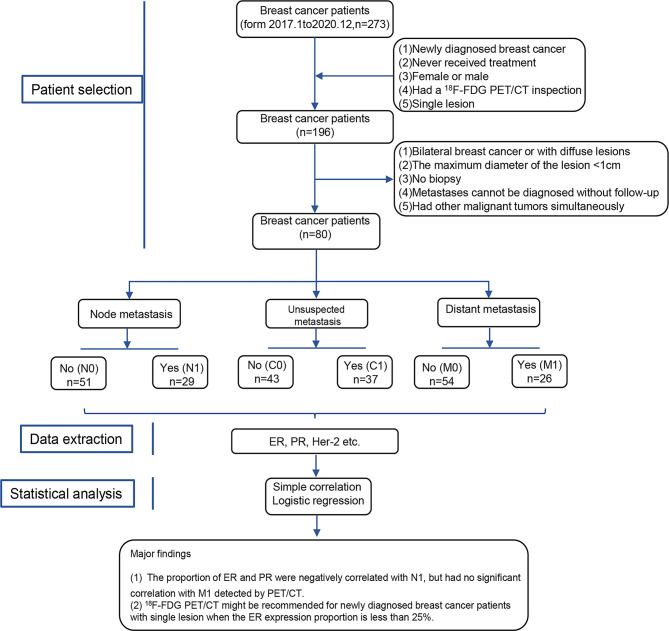
Materials and methods: Eighty newly diagnosed breast cancer patients were divided into six groups, containing N0 (no extraaxillary lymph node metastasis), N1 (extraaxillary lymph node metastasis), M0 (no distant metastasis), and M1 (distant metastasis) groups, C0 (no unsuspected metastasis), and C1 (unsuspected metastasis and treatment plan changed) detected by PET/CT. The main data, including the proportion of receptors ER (estrogen receptor), PR (progesterone receptor), and Her-2 (human epidermal growth factor receptor 2) status, were extracted. Simple correlation and logistic regression were preformed to analyze the association between them.
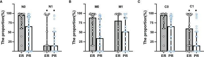
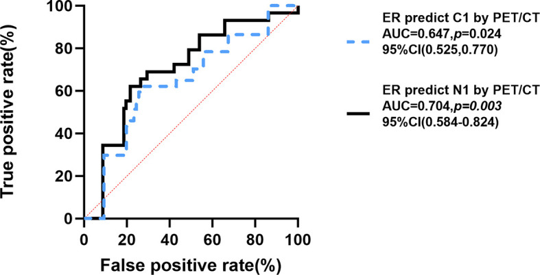
Results: Patients in N1 group had lower proportion of ER (%) and PR (%) than that in N0 group (ER: 2 [0-80] vs. 80 [15-95]; PR: 1 [0-10] vs. 20 [0-45], p<0.001). Moreover, the proportions of ER and PR were negatively correlated with N1 (ER: [r= -0.339, p= 0.002], PR: [r= -0.247, p= 0.011]) by simple correlation. Also, patients in C1 group had lower proportion of ER (%) and PR (%) than those in C0 group (ER: 10 [0-85] vs. 80 [15-90], p=0.026; PR: 1 [0-10] vs. 20 [0-70], p=0.041), while the distribution of ER and PR between M1 and M0 group had no significant difference. After the adjustment of traditional factors, the negative correlation between the proportion of ER (OR=0.986, 95% CI of OR [0.972-0.999], p=0.016) and C1 was found by logistic regression, cutoff value was 25% (ER) calculated by ROC (Receiver Operating Characteristic) curve (AUC [Area Under Curve]= 0.647, p=0.024).
Conclusion: The proportion of ER in newly diagnosed breast cancer was negatively correlated with unsuspected metastasis detected by 18F-FDG PET/CT. 18F-FDG PET/CT might be recommended for newly diagnosed breast cancer patients with single lesions when the ER expression proportion is less than 25% to find unsuspected metastasis lesions and to modify treatment plan contrasted with conventional imaging and clinical examination.
Keywords: 18F-FDG PET/CT; breast cancer; receptor status; the proportion of ER expression; the proportion of PR expression.
Copyright © 2021 Liu, Sun, Yin, Li, Liu, Liu, Zhang, Hu, Wan and Zhang.
Conflict of interest statement
The authors declare that the research was conducted in the absence of any commercial or financial relationships that could be construed as a potential conflict of interest.
Figures

Similar articles
-
18F-FDG-PET/CT for systemic staging of patients with newly diagnosed ER-positive and HER2-positive breast cancer.Eur J Nucl Med Mol Imaging. 2017 Aug;44(9):1420-1427. doi: 10.1007/s00259-017-3709-1. Epub 2017 Apr 29. Eur J Nucl Med Mol Imaging. 2017. PMID: 28456837
-
Correlation of breast cancer subtypes, based on estrogen receptor, progesterone receptor, and HER2, with functional imaging parameters from ⁶⁸Ga-RGD PET/CT and ¹⁸F-FDG PET/CT.Eur J Nucl Med Mol Imaging. 2014 Aug;41(8):1534-43. doi: 10.1007/s00259-014-2744-4. Epub 2014 Mar 21. Eur J Nucl Med Mol Imaging. 2014. PMID: 24652232
-
18F-FES PET/CT Influences the Staging and Management of Patients with Newly Diagnosed Estrogen Receptor-Positive Breast Cancer: A Retrospective Comparative Study with 18F-FDG PET/CT.Oncologist. 2019 Dec;24(12):e1277-e1285. doi: 10.1634/theoncologist.2019-0096. Epub 2019 Jul 23. Oncologist. 2019. PMID: 31337657 Free PMC article.
-
The correlation of 18F-FDG PET/CT metabolic parameters, clinicopathological factors, and prognosis in breast cancer.Clin Transl Oncol. 2021 Mar;23(3):620-627. doi: 10.1007/s12094-020-02457-w. Epub 2020 Jul 18. Clin Transl Oncol. 2021. PMID: 32683540
-
Diagnostic value of 68 Ga-DOTATATE PET-CT imaging for staging of ER+ /PR+ HER2- breast cancer patients with metastatic disease: Comparison with conventional imaging with bone scan, diagnostic CT and 18 F-FDG PET-CT in a prospective pilot trial.J Med Imaging Radiat Oncol. 2022 Sep;66(6):731-737. doi: 10.1111/1754-9485.13342. Epub 2021 Oct 21. J Med Imaging Radiat Oncol. 2022. PMID: 34676675
Cited by
-
Exploring the efficacy of 18F-FDG PET/CT in hepatocellular carcinoma diagnosis: role of Ki-67 index and tumor differentiation.Abdom Radiol (NY). 2023 Nov;48(11):3408-3419. doi: 10.1007/s00261-023-04027-4. Epub 2023 Sep 8. Abdom Radiol (NY). 2023. PMID: 37682282 Free PMC article.
References
-
- International Agency for Research on Cancer (IARC) of the World Health Organization (WHO) . World Cancer Report 2020.WHO Website. Available at: www.who.int (Accessed January 5,2021).
-
- Ayala de la Peña F, Andrés R, Garcia-Sáenz JA, Manso L, Margelí M, Dalmau E, et al. . SEOM Clinical Guidelines in Early Stage Breast Cancer (2018). Clin Trans Oncol Off Publ Fed Spanish Oncol Societies Natl Cancer Institute Mexico (2019) 21(1):18–30. doi: 10.1007/s12094-018-1973-6 - DOI - PMC - PubMed
-
- Caresia Aroztegui AP, García Vicente AM, Alvarez Ruiz S, Delgado Bolton RC, Orcajo Rincon J, Garcia Garzon JR, et al. . 18f-FDG PET/CT in Breast Cancer: Evidence-Based Recommendations in Initial Staging. Tumour Biol J Int Soc Oncodevelopmental Biol Med (2017) 39(10):1010428317728285. doi: 10.1177/1010428317728285 - DOI - PubMed
LinkOut - more resources
Full Text Sources
Research Materials
Miscellaneous