. 2021 Nov 5:11:761045.
doi: 10.3389/fonc.2021.761045. eCollection 2021.

Novel Nanoparticle-Based Cancer Treatment, Effectively Inhibits Lung Metastases and Improves Survival in a Murine Breast Cancer Model

Affiliations

Novel Nanoparticle-Based Cancer Treatment, Effectively Inhibits Lung Metastases and Improves Survival in a Murine Breast Cancer Model

Sarah Kraus et al. Front Oncol. .

Abstract

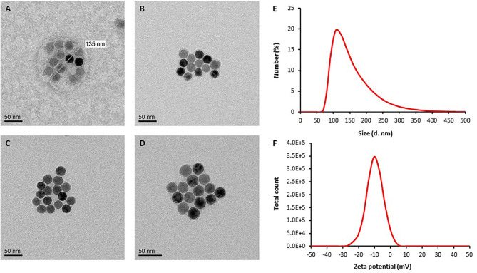
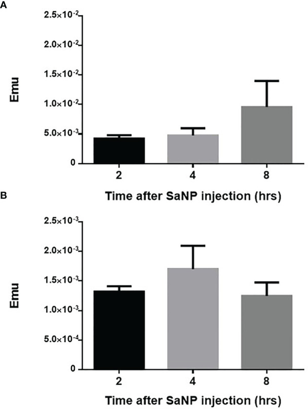
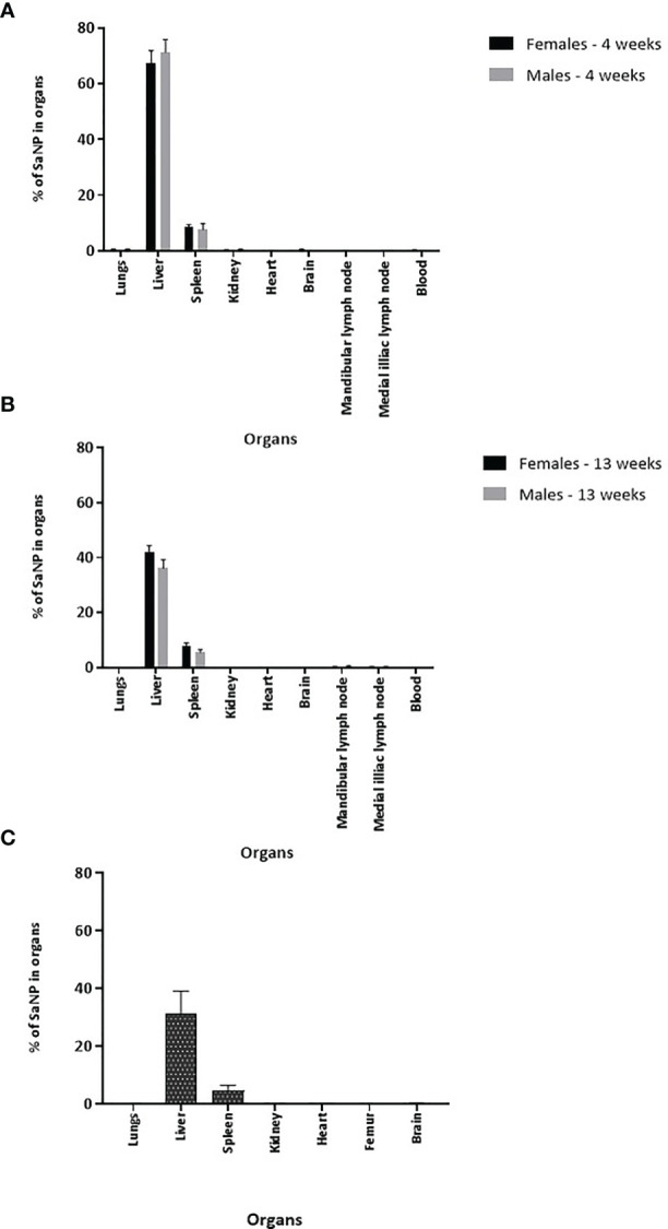
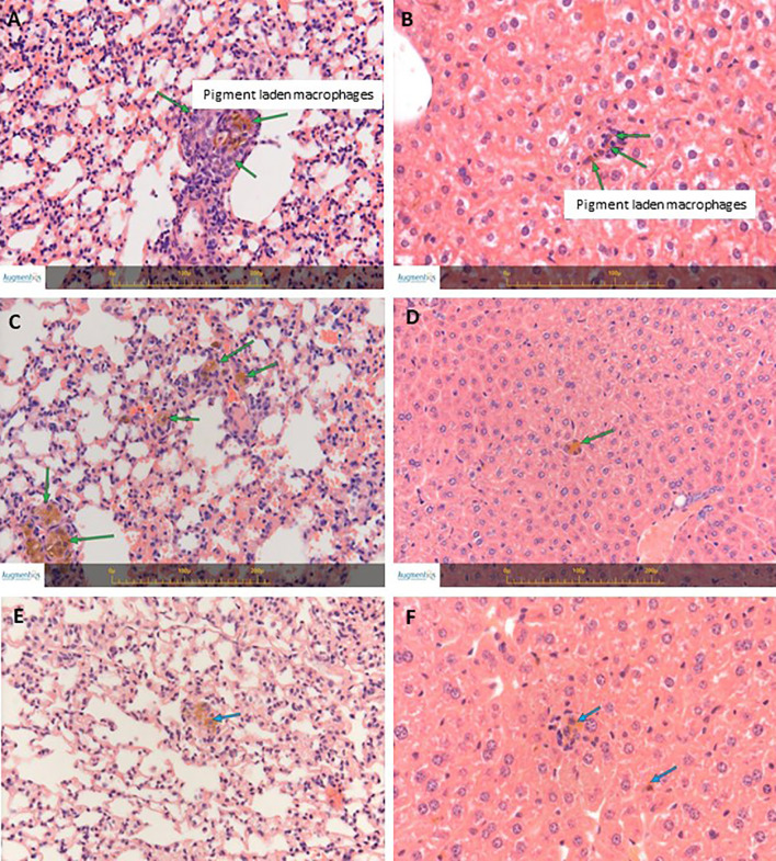
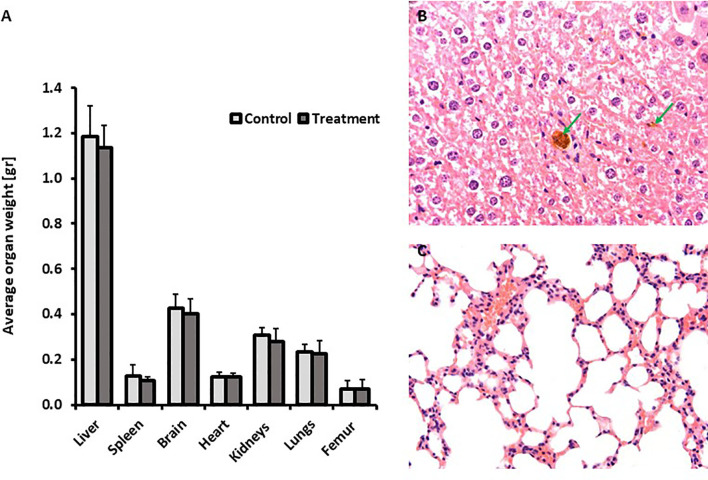
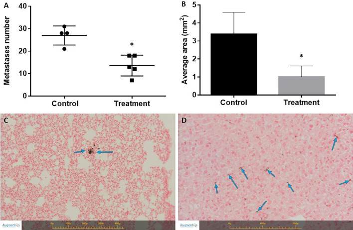
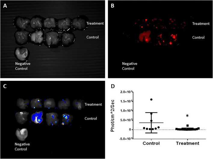
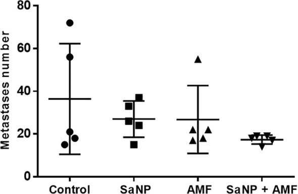
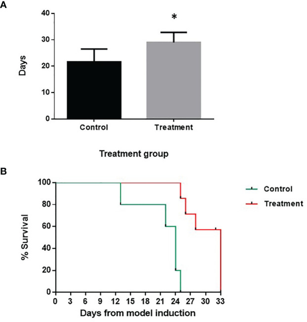
Sarah Nanoparticles (SaNPs) are unique multicore iron oxide-based nanoparticles, developed for the treatment of advanced cancer, following standard care, through the selective delivery of thermal energy to malignant cells upon exposure to an alternating magnetic field. For their therapeutic effect, SaNPs need to accumulate in the tumor. Since the potential accumulation and associated toxicity in normal tissues are an important risk consideration, biodistribution and toxicity were assessed in naïve BALB/c mice. Therapeutic efficacy and the effect on survival were investigated in the 4T1 murine model of metastatic breast cancer. Toxicity evaluation at various timepoints did not reveal any abnormal clinical signs, evidence of alterations in organ function, nor histopathologic adverse target organ toxicity, even after a follow up period of 25 weeks, confirming the safety of SaNP use. The biodistribution evaluation, following SaNP administration, indicated that SaNPs accumulate mainly in the liver and spleen. A comprehensive pharmacokinetics evaluation, demonstrated that the total percentage of SaNPs that accumulated in the blood and vital organs was ~78%, 46%, and 36% after 4, 13, and 25 weeks, respectively, suggesting a time-dependent clearance from the body. Efficacy studies in mice bearing 4T1 metastatic tumors revealed a 49.6% and 70% reduction in the number of lung metastases and their relative size, respectively, in treated vs. control mice, accompanied by a decrease in tumor cell viability in response to treatment. Moreover, SaNP treatment followed by alternating magnetic field exposure significantly improved the survival rate of treated mice compared to the controls. The median survival time was 29 ± 3.8 days in the treated group vs. 21.6 ± 4.9 days in the control, p-value 0.029. These assessments open new avenues for generating SaNPs and alternating magnetic field application as a potential novel therapeutic modality for metastatic cancer patients.

Keywords: alternating magnetic field (AMF); enhanced permeability and retention (EPR) effect; iron oxide nanoparticles; magnetic hyperthermia (MHT); metastatic breast cancer (BC).


Conflict of interest statement

Authors SK, RKD, RH, ES, ME, PR, RR, RK, AA, OS, and MC-E were employed by New Phase Ltd. The remaining authors declare that the research was conducted in the absence of any commercial or financial relationships that could be construed as a potential conflict of interest. The authors declare that this study received funding from New Phase Ltd. The funder had the following involvement with the study: Study design, collection, analysis, interpretation of data, and the writing of this article.

Figures

Figure 1
Figure 1
Representative TEM images of SaNPs. (A) SaNP with spherical shape. (B–D) SaNPs with amorphous (non-spherical) shape. Images were obtained using a Tecnai G2Spirit Twin T-12 electron microscope with an accelerating voltage of 120 kV. (E) Size (diameter) distribution of SaNPs (d. nm) determined by DLS. (F) Distribution of zeta potential values determined by DLS (mV).
Figure 2
Figure 2
Biodistribution of SaNPs in the lung metastases and blood. (A) SaNP accumulation in the lungs of BALB/c mice-bearing 4T1 metastases at 2, 4, and 8hrs post injection. (B) SaNP accumulation in the blood at 2, 4, and 8hrs post injection. Samples were analyzed by SQUID for the quantitation of IO content. Results are expressed as mean ± S.D. electromagnetic units (EMU).
Figure 3
Figure 3
Long-term tissue biodistribution of SaNPs. (A) Residual SaNP in the organs and blood of BALB/c mice at 4 weeks after a single treatment. (B) Residual SaNP at 13 weeks after a single treatment. (C) Residual SaNP at 25 weeks after 3 repeated treatments. Samples were analyzed by pEPR for the quantitation of IO content. Results are expressed as the percentage of SaNP in the various organs and blood normalized per mg/tissue.
Figure 4
Figure 4
H&E staining of lungs and livers from naïve BALB/c treated mice. (A) Lungs’ section from a mouse at 3 days post treatment. (B) Liver section from a mouse at 3 days post treatment. (C) Lungs’ section from a mouse at 14 days post treatment. (D) Liver section from a mouse at 14 days post treatment. (E) Lungs’ section from a mouse at 30 days post treatment. (F) Liver section from a mouse at 30 days post treatment. Arrows indicate small collections of pigment laden macrophages in the liver (Kupffer cells) and in the lungs, associated with minimal mononuclear cell infiltration. Images were captured using the Augmentiqs system software (14). Results are representative of 3 different experiments.
Figure 5
Figure 5
Repeated dose chronic toxicity study. (A) Average organ weight after a follow up period of 25 weeks. (B) Representative liver section (H&E staining) from a treated mouse at 25 weeks post treatment. Green arrows indicate pigment laden macrophages in the liver (Kupffer cells) associated with minimal mononuclear cell infiltration. (C) Representative lungs’ section (H&E staining) from a treated mouse at 25 weeks post treatment. No changes were observed. Images were captured using the Augmentiqs system software (14).
Figure 6
Figure 6
Effect of treatment on the number of lung metastases and tumor size in the 4T1 breast cancer model. (A) Number of lung metastases. Mice were sacrificed on day 17 following cancer model induction. The lungs were excised and the number of metastases was counted. *Statistically significant difference. (B) Tumor size determined by histopathology and 2-D morphometric analysis expressed as average tumor area (mm2). *Statistically significant difference. (C) Representative section of Prussian blue staining of the lungs of treated mice. Blue arrows indicate Prussian blue positive pigment laden macrophages. No inflammation was associated with the presence of pigment. (D) Representative section of Prussian blue staining of the liver of treated mice. Blue arrows indicate Prussian blue positive pigment laden macrophages (Kupffer cells). No inflammation was associated with the presence of pigment. Images were captured using the Augmentiqs system software (14).
Figure 7
Figure 7
CRi Maestro fluorescent ex-vivo imaging of the lungs. (A) Lungs – grayscale. (B) Fluorescent imaging of mCherry metastases. (C) Heat map of viable metastases. (D) Quantitation of total fluorescent signal in the lungs per group, expressed in x106phot/cm2/sec. Each dot represents the fluorescence intensity of individual metastases. Normal lungs without mCherry expression were used as a negative control. Each pair of lungs may contain several metastases that express different fluorescence intensities. Results are representative of 3 different experiments. *Statistically significant difference.
Figure 8
Figure 8
Effect of treatment on the number of lung metastases in the 4T1 breast cancer model. Treatment cycles started at day 14 after cell inoculation. Mice were sacrificed on day 21 post treatment. The lungs were excised and the number of metastases was counted.
Figure 9
Figure 9
Effect of treatment on animal survival in the 4T1 breast cancer lung metastasis model. Treatment cycles started at day 13 after cell inoculation. (A) Median survival time (days) of control vs. treatment group. (B) Kaplan-Meier survival curve. There was a significant difference in the survival rate between the two groups. The statistical analysis was performed using the log-rank test, p-value <0.005 (*).

References

    1. Siegel RL, Miller KD, Fuchs HE, Jemal A. Cancer Statistics, 2021. CA Cancer J Clin (2021) 71:7–33. doi: 10.3322/caac.21654 - DOI - PubMed
    1. Azarmi S, Roa WH, Lobenberg R. Targeted Delivery of Nanoparticles for the Treatment of Lung Diseases. Adv Drug Deliv Rev (2008) 60:863–75. doi: 10.1016/j.addr.2007.11.006 - DOI - PubMed
    1. Jain S, Hirst DG, O'Sullivan JM. Gold Nanoparticles as Novel Agents for Cancer Therapy. Br J Radiol (2012) 85:101–13. doi: 10.1259/bjr/59448833 - DOI - PMC - PubMed
    1. Wust P, Hildebrandt B, Sreenivasa G, Rau B, Gellermann J, Riess H, et al. . Hyperthermia in Combined Treatment of Cancer. Lancet Oncol (2002) 3:487–97. doi: 10.1016/S1470-2045(02)00818-5 - DOI - PubMed
    1. Rabinovitz R, Shalev O, Kraus S. Novel Approach for Magnetic Nanoparticle-Based Hyperthermia for Metastatic Cancer Treatment. BioMed J Sci Tech Res (2020) 32:25025–27. doi: 10.26717/BJSTR.2020.32.005254 - DOI

LinkOut - more resources