

Transcriptome Profile During Rabies Virus Infection: Identification of Human CXCL16 as a Potential New Viral Target
- PMID: 34804996
- PMCID: PMC8602097
- DOI: 10.3389/fcimb.2021.761074
Transcriptome Profile During Rabies Virus Infection: Identification of Human CXCL16 as a Potential New Viral Target
Abstract
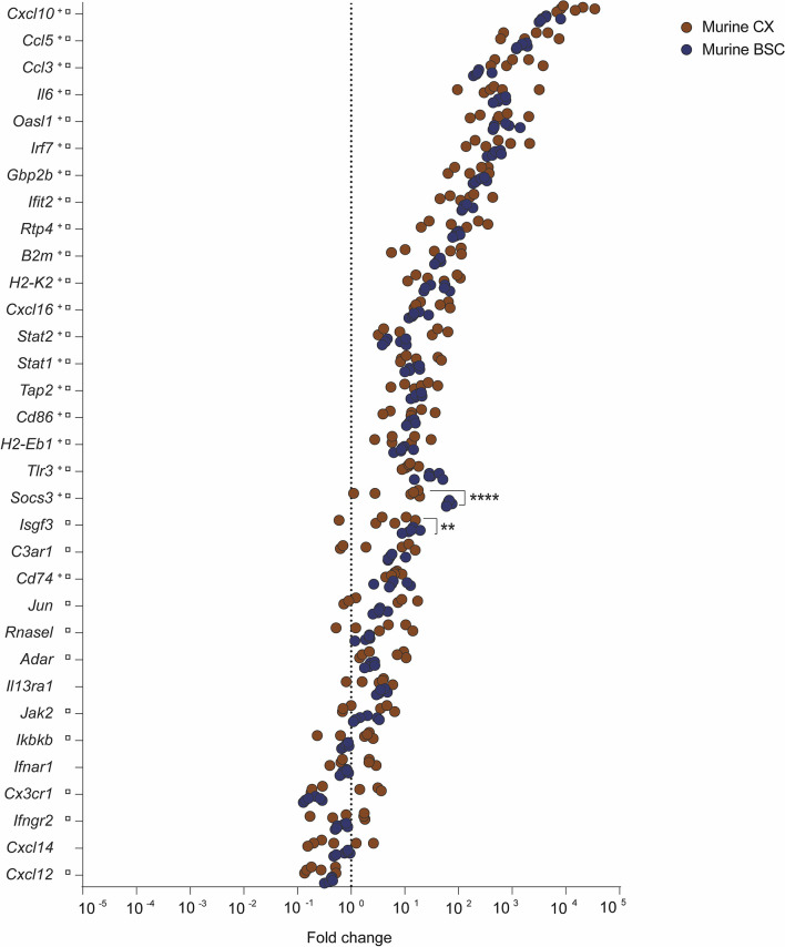
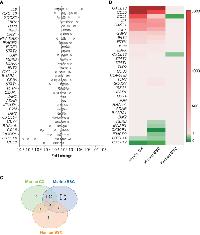
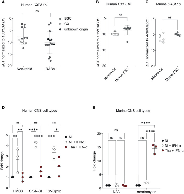
Rabies virus (RABV), the causative agent for rabies disease is still presenting a major public health concern causing approximately 60,000 deaths annually. This neurotropic virus (genus Lyssavirus, family Rhabdoviridae) induces an acute and almost always fatal form of encephalomyelitis in humans. Despite the lethal consequences associated with clinical symptoms of rabies, RABV limits neuro-inflammation without causing major histopathological lesions in humans. Nevertheless, information about the mechanisms of infection and cellular response in the central nervous system (CNS) remain scarce. Here, we investigated the expression of inflammatory genes involved in immune response to RABV (dog-adapted strain Tha) in mice, the most common animal model used to study rabies. To better elucidate the pathophysiological mechanisms during natural RABV infection, we compared the inflammatory transcriptome profile observed at the late stage of infection in the mouse brain (cortex and brain stem/cerebellum) with the ortholog gene expression in post-mortem brain biopsies of rabid patients. Our data indicate that the inflammatory response associated with rabies is more pronounced in the murine brain compared to the human brain. In contrast to murine transcription profiles, we identified CXC motif chemokine ligand 16 (CXCL16) as the only significant differentially expressed gene in post-mortem brains of rabid patients. This result was confirmed in vitro, in which Tha suppressed interferon alpha (IFN-α)-induced CXCL16 expression in human CNS cell lines but induced CXCL16 expression in IFN-α-stimulated murine astrocytes. We hypothesize that RABV-induced modulation of the CXCL16 pathway in the brain possibly affects neurotransmission, natural killer (NK) and T cell recruitment and activation. Overall, we show species-specific differences in the inflammatory response of the brain, highlighted the importance of understanding the potential limitations of extrapolating data from animal models to humans.
Keywords: CXCL16; brain; human; immune response; mouse; neuroinflammation; rabies; transcriptome.
Copyright © 2021 Feige, Sáenz-de-Santa-María, Regnault, Lavenir, Lepelletier, Halacu, Rajerison, Diop, Nareth, Reynes, Buchy, Bourhy and Dacheux.
Conflict of interest statement
Author PB is employed by GSK Vaccines. The remaining authors declare that the research was conducted in the absence of any commercial or financial relationships that could be construed as a potential conflict of interest.
Figures
Similar articles
-
Inflammatory response and MAPK and NF-κB pathway activation induced by natural street rabies virus infection in the brain tissues of dogs and humans.Virol J. 2020 Oct 20;17(1):157. doi: 10.1186/s12985-020-01429-4. Virol J. 2020. PMID: 33081802 Free PMC article.
-
Interferon-λ Attenuates Rabies Virus Infection by Inducing Interferon-Stimulated Genes and Alleviating Neurological Inflammation.Viruses. 2020 Apr 6;12(4):405. doi: 10.3390/v12040405. Viruses. 2020. PMID: 32268591 Free PMC article.
-
Immunological findings of West Caucasian bat virus in an accidental host.J Virol. 2025 Feb 25;99(2):e0191424. doi: 10.1128/jvi.01914-24. Epub 2025 Jan 23. J Virol. 2025. PMID: 39846740 Free PMC article.
-
Role of chemokines in rabies pathogenesis and protection.Adv Virus Res. 2011;79:73-89. doi: 10.1016/B978-0-12-387040-7.00005-6. Adv Virus Res. 2011. PMID: 21601043 Free PMC article. Review.
-
Regulation of innate immune responses by rabies virus.Animal Model Exp Med. 2022 Oct;5(5):418-429. doi: 10.1002/ame2.12273. Epub 2022 Sep 22. Animal Model Exp Med. 2022. PMID: 36138548 Free PMC article. Review.
Cited by
-
Antiviral mechanisms of two broad-spectrum monoclonal antibodies for rabies prophylaxis and therapy.Front Immunol. 2023 Aug 10;14:1186063. doi: 10.3389/fimmu.2023.1186063. eCollection 2023. Front Immunol. 2023. PMID: 37638057 Free PMC article.
-
Tracking lethal threat: in-depth review of rabies.Open Vet J. 2023 Nov;13(11):1385-1399. doi: 10.5455/OVJ.2023.v13.i11.1. Epub 2023 Nov 30. Open Vet J. 2023. PMID: 38107233 Free PMC article. Review.
-
Viral sepsis - pathophysiology and disease manifestation.Infection. 2025 Jun;53(3):775-784. doi: 10.1007/s15010-025-02486-z. Epub 2025 Feb 17. Infection. 2025. PMID: 39961996 Free PMC article. Review.
-
Hydrogel-based 3D human iPSC-derived neuronal culture for the study of rabies virus infection.Front Cell Infect Microbiol. 2023 Aug 25;13:1215205. doi: 10.3389/fcimb.2023.1215205. eCollection 2023. Front Cell Infect Microbiol. 2023. PMID: 37692167 Free PMC article.
-
A One Medicine Mission for an Effective Rabies Therapy.Front Vet Sci. 2022 Mar 16;9:867382. doi: 10.3389/fvets.2022.867382. eCollection 2022. Front Vet Sci. 2022. PMID: 35372555 Free PMC article.
References
-
- Baer G. M. (1975). The Natural History of Rabies, CRC Press (New York: Academic Press; ), 640.
Publication types
MeSH terms
Substances
LinkOut - more resources
Full Text Sources
Medical