

Myeloid sarcoma: an uncommon presentation of myeloid neoplasms; a case series of 4 rare cases reported in a tertiary care institute
- PMID: 34805008
- PMCID: PMC8597781
- DOI: 10.4322/acr.2021.339
Myeloid sarcoma: an uncommon presentation of myeloid neoplasms; a case series of 4 rare cases reported in a tertiary care institute
Abstract
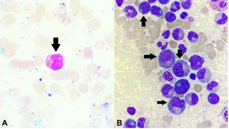
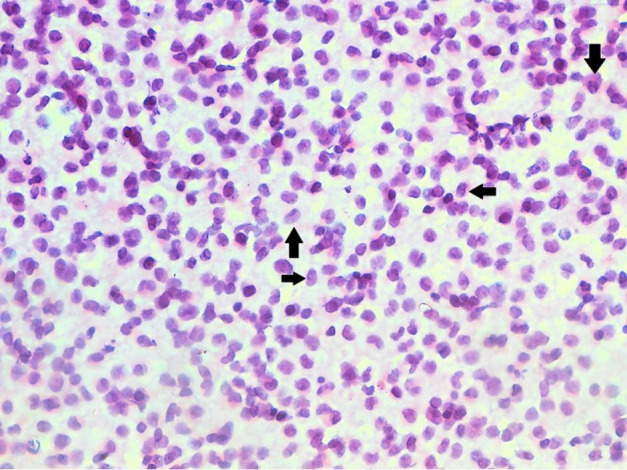
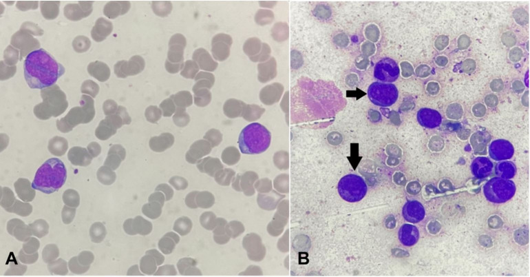
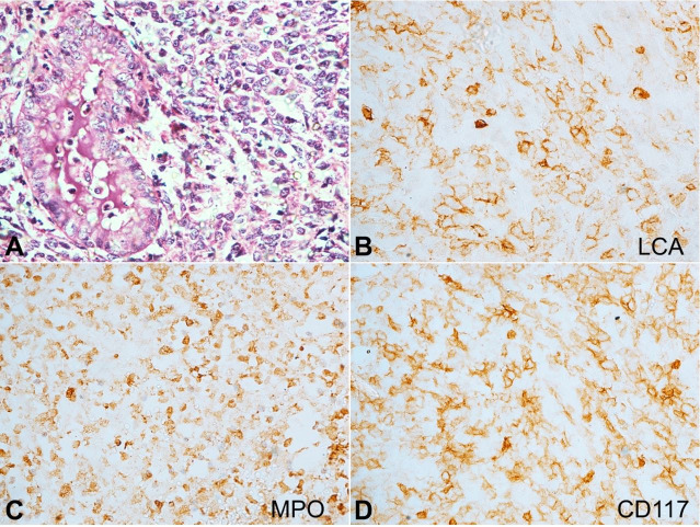
Myeloid sarcoma (MS) is a rare extramedullary neoplasm of myeloid cells, which can arise before, concurrently with, or following hematolymphoid malignancies. We report 04 such cases of MS, diagnosed in this institute over a period of 6 years, during various phases of their respective myeloid neoplasms/leukemias. These cases include MS occurring as a relapse of AML (Case 1), MS occurring as an initial presentation of CML (Case 2), MS occurring during ongoing chemotherapy in APML (Case 3), and MS presenting as a progression of MDS to AML (Case 4). In the absence of relevant clinical history and unemployment of appropriate immunohistochemical (IHC) studies, these cases have a high risk of being frequently misdiagnosed either as Non-Hodgkin's Lymphoma (NHL) or small round cell tumors or undifferentiated carcinomas, which may further delay their management, making an already bad prognosis worse. This case series has been designed to throw light on the varied presentation of MS and the lineage differentiation of its neoplastic cells through the application of relevant IHC markers along with their clinical correlation.
Keywords: Leukemia, Myelogenous, Chronic, BCR-ABL Positive; Leukemia, Myeloid, Acute; Leukemia, Promyelocytic, Acute; Myelodysplastic Syndromes; Sarcoma, Myeloid.
Copyright: © 2021 The Authors.
Conflict of interest statement
Conflict of interest: None.
Figures
Similar articles
-
Myeloid sarcoma: Experience from a tertiary care center in southern India - A series of eight unusual cases.Indian J Pathol Microbiol. 2025 Jan 1;68(1):113-117. doi: 10.4103/ijpm.ijpm_474_23. Epub 2024 Jun 20. Indian J Pathol Microbiol. 2025. PMID: 38904435
-
Ph-negative non-Hodgkin's lymphoma occurring in chronic phase of Ph-positive chronic myelogenous leukemia is defined as a genetically different neoplasm from extramedullary localized blast crisis: report of two cases and review of the literature.Leukemia. 2000 Jan;14(1):169-82. doi: 10.1038/sj.leu.2401606. Leukemia. 2000. PMID: 10637493 Review.
-
Myeloid sarcoma of the small intestine in nonleukemic patients - A report of three cases with review of literature.Indian J Cancer. 2024 Oct 1;61(4):676-686. doi: 10.4103/ijc.IJC_1055_20. Epub 2025 Feb 17. Indian J Cancer. 2024. PMID: 39960694 Review.
-
Minimally differentiated acute myelogenous leukemia (AML-M0) granulocytic sarcoma presenting in the oral cavity.Oral Oncol. 2002 Jul;38(5):516-9. doi: 10.1016/s1368-8375(01)00085-9. Oral Oncol. 2002. PMID: 12110349
-
Extramedullary myeloid cell tumors in myelodysplastic-syndromes: not a true indication of impending acute myeloid leukemia.Leuk Lymphoma. 1996 Mar;21(1-2):153-9. doi: 10.3109/10428199609067593. Leuk Lymphoma. 1996. PMID: 8907283 Review.
Cited by
-
An Overview of Advances in Rare Cancer Diagnosis and Treatment.Int J Mol Sci. 2024 Jan 18;25(2):1201. doi: 10.3390/ijms25021201. Int J Mol Sci. 2024. PMID: 38256274 Free PMC article. Review.
-
Anatomic Approach to Common and Uncommon Manifestations of Thoracic Leukemias with Radiologic-Pathologic Correlation.Radiol Cardiothorac Imaging. 2023 Dec;5(6):e230151. doi: 10.1148/ryct.230151. Radiol Cardiothorac Imaging. 2023. PMID: 38166347 Free PMC article.
-
Spinal myeloid sarcoma presenting as initial symptom in acute promyelocytic leukemia with a rare cryptic PLZF::RARα fusion gene: a case report and literature review.Front Oncol. 2024 May 21;14:1375737. doi: 10.3389/fonc.2024.1375737. eCollection 2024. Front Oncol. 2024. PMID: 38835381 Free PMC article.
References
-
- Baveja P, Jadhav T, Pendkur G. Granulocytic sarcoma presenting as a palpable breast lump in a 13-year-old female as a relapse of AML. Indian J Pathol Oncol. 2020;7(4):669–673. doi: 10.18231/j.ijpo.2020.132. - DOI
-
- Rappaport H. In: Atlas of tumor pathology. Armed Forces Institute of Pathology, editor. Washington: AFIP; 1966. Tumors of the Hematopoietic System.
Publication types
LinkOut - more resources
Full Text Sources
Research Materials
Miscellaneous
