. 2021 Nov 11:2021:3862492.
doi: 10.1155/2021/3862492. eCollection 2021.

Duck Interleukin-22: Identification and Expression Analysis in Riemerella anatipestifer Infection

Affiliations

Duck Interleukin-22: Identification and Expression Analysis in Riemerella anatipestifer Infection

Rochelle A Flores et al. J Immunol Res. .

Abstract

Riemerella anatipestifer is one of the most devastating pathogens affecting the global duck farms. Infection is involved in secretion of proinflammatory cytokines, including interleukin- (IL-) 17A. During the immune response to infection, IL-22 and IL-17A are often produced concurrently and at high levels in inflamed tissues. Little is known about duck IL-22 (duIL-22) during R. anatipestifer infection. We describe the characterization of duIL-22 and its mRNA expression analysis in splenic lymphocytes and macrophages treated with heat-killed R. anatipestifer and in the spleens and livers of R. anatipestifer-infected ducks. Full-length cDNA of duIL-22 encoded 197 amino acids. The deduced amino acid sequence of duIL-22 shared a 30.4-40.5% similarity with piscine counterparts, 57.4-60.1% with mammalian homologs, and 93.4% similarity to the chicken. Duck IL-22 mRNA expression level was relatively high in the skin of normal ducks. It was increased in mitogen-stimulated splenic lymphocytes and in killed R. anatipestifer-activated splenic lymphocytes and macrophages. Compared with healthy ducks, IL-22 transcript expression was significantly upregulated in the livers and spleens on days 1 and 4 postinfection, but not on day 7. IL-17A was significantly increased in the spleens only on day 4 postinfection and in the livers at all time points. When splenic lymphocytes were stimulated with heat-killed R. anatipestifer, CD4+ cells predominantly produced IL-22 while IL-17A was expressed both by CD4+ and CD4- cells. These results suggested that IL-22 and IL-17A are likely expressed in different cell types during R. anatipestifer infection.


Conflict of interest statement

The authors declare that they have no conflicts of interest.

Figures
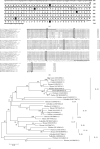
Figure 1
Figure 1
Molecular characterization and genetic analysis of duck IL-22 cDNA. (a) Sequences of nucleotide and deduced amino acid of duck IL-22. The underline indicated the predicted signal peptide region, black boxes indicated the conserved cysteine residues, and white box indicated the putative N-linked glycosylation area. (b) Multiple alignment of amino acid sequence of avian and mammalian IL-22. Clustal Omega software was used for multiple alignments. The identical residues among sequences were indicated with asterisks (∗). The underline indicated the signal peptide of duck IL-22, white boxes indicated the conserved cysteine residues between species, and the conserved IL-10 family motif is underlined using a dashed line. GenBank accession numbers used in the comparison are written after the species names. (c) Phylogenetic tree indicating the relationships between IL-22 amino acid sequences and other known IL-10 family members. The tree was made using the neighbor-joining method (MEGA version 7 program) and amino acid multiple alignments. The node values indicated percentage bootstrap confidence levels obtained from ten thousand replicates. The sequence accession numbers are written after the species name. The white box indicated duck IL-22.
Figure 2
Figure 2
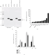
Molecular weight of duck IL-22 and its expression in normal tissues and stimulated splenic lymphocytes. (a) Western blot analysis to determine molecular weight of duck IL-22 protein from COS-7 cells transfected with duIL-22-MYC construct. Supernatants and cell lysates of COS-7 cells were obtained at 48 h after transfection and deglycosylated using PNGase F (100 U peptide-N-glycosidase F) at 37°C for 1 h. Supernatants and cell lysates under reducing conditions were separated using SDS-PAGE. An anti-MYC antibody was used to detect the specific bands of duck IL-22. The asterisk (∗) and arrow indicated the normal bands and the deglycosylated bands of duck IL-22, respectively. (b) Distribution of IL-22 transcripts in normal healthy duck tissues. Total RNA was obtained from tissues of two-week-old normal ducks (n = 5) and used to qRT-PCR analysis. Expression levels of β-actin were used for normalization of IL-22 expression levels, which were calibrated to the lowest expression level detected. Results are presented as the mean ± SE values from 2 independent experiments. (c) Duck IL-22 expression levels in mitogen-treated splenic lymphocytes. The lymphocytes were obtained from two-week-old normal ducks using Ficoll density gradient centrifugation. They were activated using 25 μg/ml poly I:C, 10 μg/m LPS, or 10 μg/ml ConA for the indicated times. Expression levels of β-actin gene were used for normalization of IL-22 expression level, which were calibrated with untreated cultured splenic lymphocytes (NC). Results are presented as the mean ± SE values from 2 independent experiments performed in triplicate. P < 0.05 and ∗∗∗P < 0.001.
Figure 3
Figure 3
mRNA expression profiles of duck IL-22 in splenic lymphocytes and macrophages. Lymphocytes and macrophages were collected and isolated from 2-week-old healthy ducks and treated with heat-inactivated R. anatipestifer serotype 7 for the indicated times. RNA was isolated and samples were applied to qRT-PCR analysis. The transcript expression levels of IL-22 (a) and IL-17A (b) were normalized to expression levels of β-actin used as a reference gene and presented relative to the expression levels of untreated/cultured lymphocytes and macrophages (NC). Results are presented as the mean ± SE values from 3 independent experiments performed in triplicate. ∗∗P < 0.01 and ∗∗∗P < 0.001. RA: R. anatipestifer-stimulated lymphocytes.
Figure 4
Figure 4
Expression profiles of IL-22 in ducks infected with R. anatipestifer. Expression levels of IL-22 (a) and IL-17A (b) transcripts from the spleens and livers of ducks infected intramuscularly with 5 × 10⁷ CFUs of R. anatipestifer serotype 7. The spleens and livers of 2-week-old healthy ducks were aseptically sampled on days 1, 4, and 7 postinfection (dpi). Tissue samples were pooled, and total RNA was isolated for qRT-PCR. Gene expression levels were normalized with β-actin used as a reference gene and presented relative to the expression levels of uninfected and healthy controls (NC). These results represented one representative of 2 independent experiments. Results are presented as the mean ± SE values (n = 5). P < 0.05 and ∗∗∗P < 0.001. dpi: days postinfection; RA: R. anatipestifer-infected ducks.
Figure 5
Figure 5
IL-22 is produced primarily by CD4+ cells during R. anatipestifer infection, but not IL-17. The spleens were collected from 2-week-old healthy ducks. Spleen cells were isolated, cultured, and stimulated with heat-killed R. anatipestifer serotype 7. After 24 h, cells were collected, labelled, and separated following incubation in MACS buffer. Total RNA was obtained, and qRT-PCR analysis was performed. Gene expression levels of IL-22 (a) and IL-17A (b) transcripts were normalized with β-actin used as a reference gene and calibrated with the expression levels of untreated cultured spleen cells (NC). These results represented one representative of 2 independent experiments. Results are presented as the mean ± SE values performed in triplicate. ∗∗P < 0.01. RA: splenic lymphocytes stimulated with heat-inactivated R. anatipestifer.

Similar articles

Cited by

References

    1. Dudakov J. A., Hanash A. M., Van Den Brink M. R. M. Interleukin-22: immunobiology and pathology. Annual Review of Immunology . 2015;33:747–785. doi: 10.1146/annurev-immunol-032414-112123. - DOI - PMC - PubMed
    1. Eyerich K., Dimartino V., Cavani A. IL-17 and IL-22 in immunity: driving protection and pathology. European Journal of Immunology . 2017;47:607–614. doi: 10.1002/eji.201646723. - DOI - PubMed
    1. Ronacher K., Sinha R., Cestari M. IL-22: an underestimated player in natural resistance to tuberculosis? Frontiers in Immunology . 2018;9:1–7. doi: 10.3389/fimmu.2018.02209. - DOI - PMC - PubMed
    1. Rutz S., Eidenschenk C., Ouyang W. IL-22, not simply a Th17 cytokine. Immunological Reviews . 2013;252:116–132. doi: 10.1111/imr.12027. - DOI - PubMed
    1. Valeri M., Raffatellu M. Cytokines IL-17 and IL-22 in the host response to infection. Pathogens and Disease . 2016;74:1–15. doi: 10.1093/femspd/ftw111. - DOI - PMC - PubMed

Supplementary concepts