. 2021 Oct 25:6:59.
doi: 10.21037/tgh-20-314. eCollection 2021.

Percutaneous microwave ablation of hepatic lesions near the heart

Affiliations

Percutaneous microwave ablation of hepatic lesions near the heart

Sreeja Sanampudi et al. Transl Gastroenterol Hepatol. .

Abstract

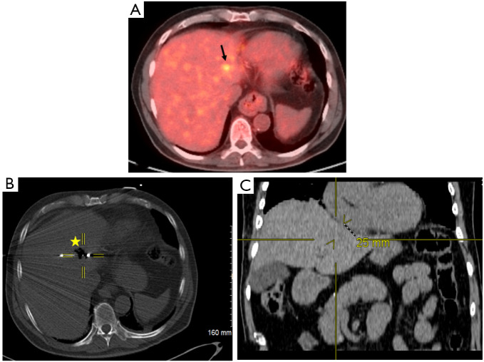
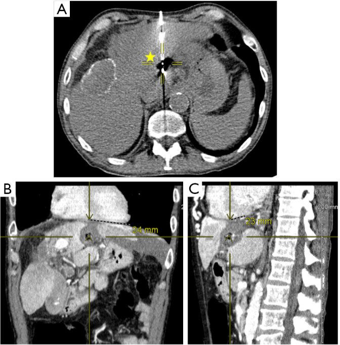
Background: Early stage liver cancer is often treated with hepatic resection or transplantation for curative intent. Microwave ablation (MWA) is often performed in patients who are poor surgical candidates, patients with limited multifocal disease, disease close to hepatic vasculature, but can also be performed with curative intent in case of small lesions. The purpose of this study is to evaluate safety and efficacy of MWA of liver tumors with final ablation zone ≤5 mm from the heart.

Methods: A retrospective review was conducted on patients with hepatic cancer who underwent MWA between 1/2015 and 6/2019. Patients with a final ablation zone ≤5 mm to the heart were included. For these patients, imaging obtained prior, during and after procedure along with procedure reports were used to identify tumor and ablation characteristics, and electronic medical records were used to identify patient demographics and disease status.

Results: A total of 17 patients had liver tumors with ablation zone ≤5 mm to the heart. Mean lesion size was 18.2 mm (range, 10-33 mm) and mean follow-up period was 10.4 months. Of note 82% of patients had multifocal disease at time of MWA of lesion close to the heart. Two patients had pneumothorax, one of which required chest tube placement. None of the patients had cardiac arrhythmias or other complications. Overall 12/17 of the patients had disease progression within the liver at different sites from ablated lesions. One patient had residual disease and one had local recurrence. In addition, 4/17 patients, had no disease progression or recurrence and one underwent liver transplantation prior to follow-up imaging.

Conclusions: MWA of liver lesions with ablation zone ≤5 mm to the heart is safe and effective, however, it can be technically challenging.

Keywords: Microwave ablation (MWA); heart; hepatic lesions; hepatocellular carcinoma (HCC); liver metastases.


Conflict of interest statement

Conflicts of Interest: All authors have completed the ICMJE uniform disclosure form (available at http://dx.doi.org/10.21037/tgh-20-314). The authors have no conflicts of interest to declare.

Figures

Figure 1
Figure 1
Localization of primary hepatocellular carcinoma and proximity to the heart. (A) Pre-procedural positron electron tomography/computed tomography (CT) scan demonstrating 11 mm hypermetabolic hepatic lesion (black arrow) in a patient with primary hepatocellular carcinoma. (B) Perioperative CT fluoroscopic imaging demonstrating 4.2 cm spherical zone of ablation (star) with crosshairs centered on the emitter of the microwave antenna. (C) Post-procedural coronal CT image that corresponds to the center of ablation zone. Yellow crosshairs were localized to the same point as center of ablation zone in this coronal view. Distance from center of ablation zone to the heart was measured. It can be noted on the coronal view the center of ablation zone to the heart measures 25 mm in radius corresponding to ≤5 mm distance from the heart.
Figure 2
Figure 2
Ablation of hepatocellular carcinoma. The following images are from a patient with 11 mm left hepatic lobe lesion that was primary hepatocellular carcinoma. (A) Peri-operative computed tomography (CT) image demonstrating 4.2 cm spherical ablation zone (yellow star) with crosshairs centered to emission point of microwave antenna. (B,C) Post ablation CT venous phase demonstrating ablation cavity in coronal (B) and sagittal (C) views. Crosshairs and distances are highlighted in coronal (B) and sagittal (C) views demonstrating a distance of 24 and 23 mm respectively between centers of ablation zone to the heart in a patient with 4.2 cm spherical ablation zone. Thus, in both coronal and sagittal views, the distance of the final ablation zone was ≤5 mm to the heart.

References

    1. Siegel RL, Miller KD, Jemal A. Cancer statistics, 2020. CA Cancer J Clin 2020;70:7-30. 10.3322/caac.21590 - DOI - PubMed
    1. American Cancer Society. Facts & Figures 2020. American Cancer Society. Atlanta, 2020. - PMC - PubMed
    1. de Ridder J, de Wilt JH, Simmer F, et al. Incidence and origin of histologically confirmed liver metastases: an explorative case-study of 23,154 patients. Oncotarget 2016;7:55368-76. 10.18632/oncotarget.10552 - DOI - PMC - PubMed
    1. Meloni MF, Chiang J, Laeseke PF, et al. Microwave ablation in primary and secondary liver tumors: technical and clinical approaches. Int J Hyperthermia 2017;33:15-24. 10.1080/02656736.2016.1209694 - DOI - PMC - PubMed
    1. Glassberg MB, Ghosh S, Clymer JW, et al. Microwave ablation compared with hepatic resection for the treatment of hepatocellular carcinoma and liver metastases: a systematic review and meta-analysis. World J Surg Oncol 2019;17:98. 10.1186/s12957-019-1632-6 - DOI - PMC - PubMed