

doi: 10.1016/j.jimed.2020.01.001.
eCollection 2020 Feb.
Application of three-dimensional printing in interventional medicine
Affiliations
- PMID: 34805900
- PMCID: PMC8562235
- DOI: 10.1016/j.jimed.2020.01.001
Item in Clipboard
Application of three-dimensional printing in interventional medicine
J Interv Med.
.
Erratum in
-
Erratum: An erratum on "Application of three-dimensional printing in interventional medicine" [J Interv Med (February 2020) 1-16].J Interv Med. 2020 Sep 19;3(3):156. doi: 10.1016/j.jimed.2020.06.001. eCollection 2020 Sep. J Interv Med. 2020. PMID: 34826305 Free PMC article.
No abstract available
Figures
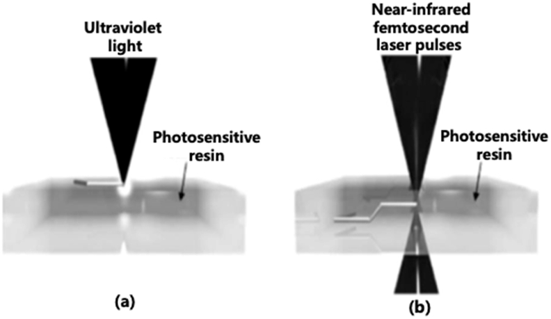
Schematic showing SLA printing at the surface of a photosensitive polymer (a) and TPP printing at the focal point inside the resin volume (b).
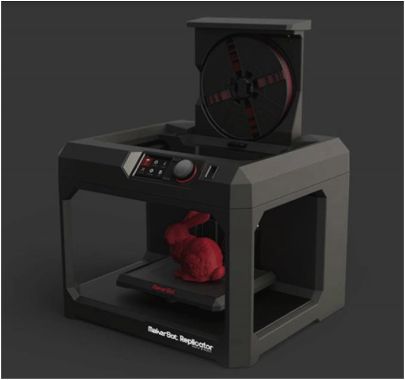
Picture showing the fused deposition modeling 3D printer (MakerBot Replicator 2 Desktop 3D Printer).
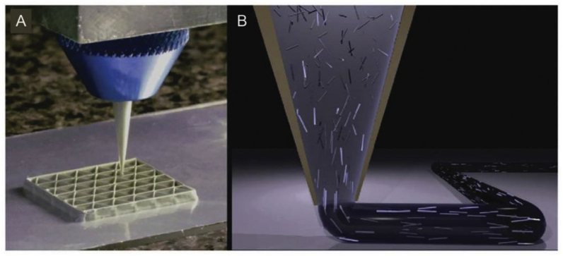
Schematic of the nozzle in DIW printer.
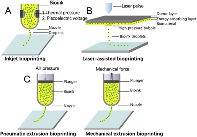
Illustrations of three mainstay bioprinting techniques. (A) IBP. (B) Laser-assisted bioprinting. (C) Extrusion bioprinting.
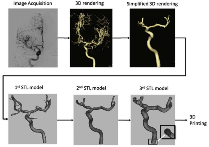
Preparation steps for 3DP.
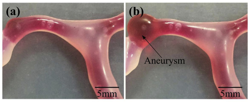
Comparison of the brain vascular model. (a) No blister-like dilation bulges in the vascular model; (b) presence of a blister-like aneurysm.
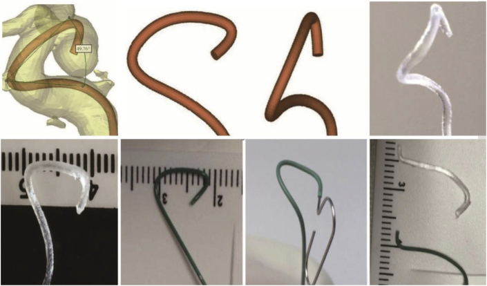
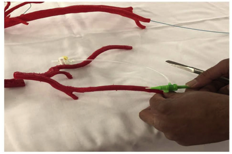
Pre-planning of the shaping mandrel of microcatheter using 3DP rapid prototyping technology.
Application of 3DP in selecting the best-shaped microcatheter tips using a 3D software, and the tips were printed using 3D printers. Using this method, it was possible to enhance the accuracy and stability of the microcatheter position during embolization.
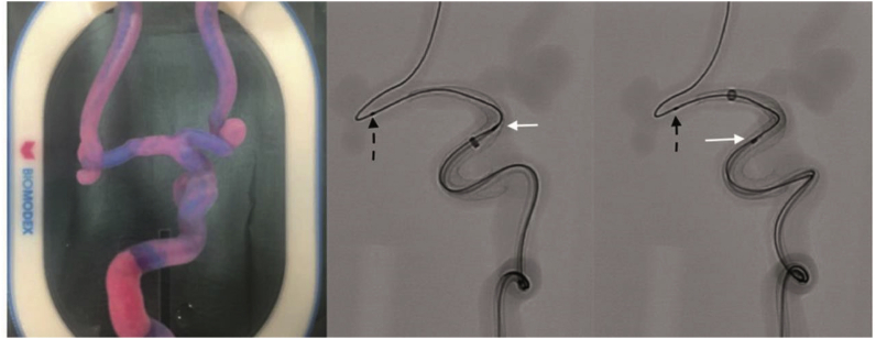
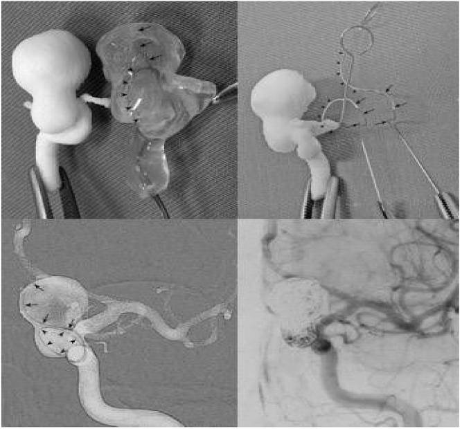
Surgical simulation of flow diverting treatment of intracranial anterior circulation aneurysm with 3DP silicone replica. White arrows indicate pipeline shield and black dashed arrows indicate the microwire.
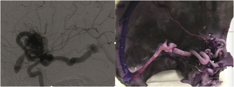
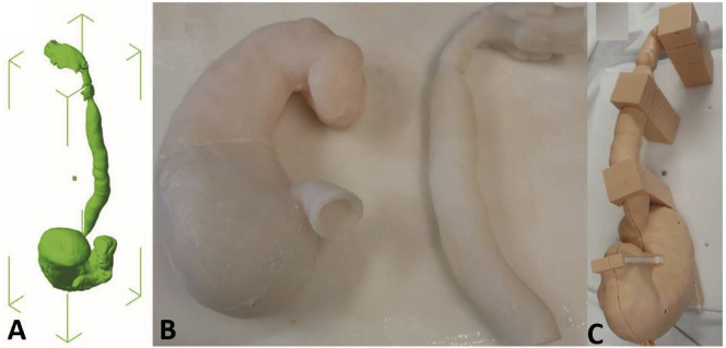
(a) A case of a 21-year old male patient with Sylvian fissure bAVM (b) and printed 3D model showing the nidus and the arterial feeders and the draining veins.
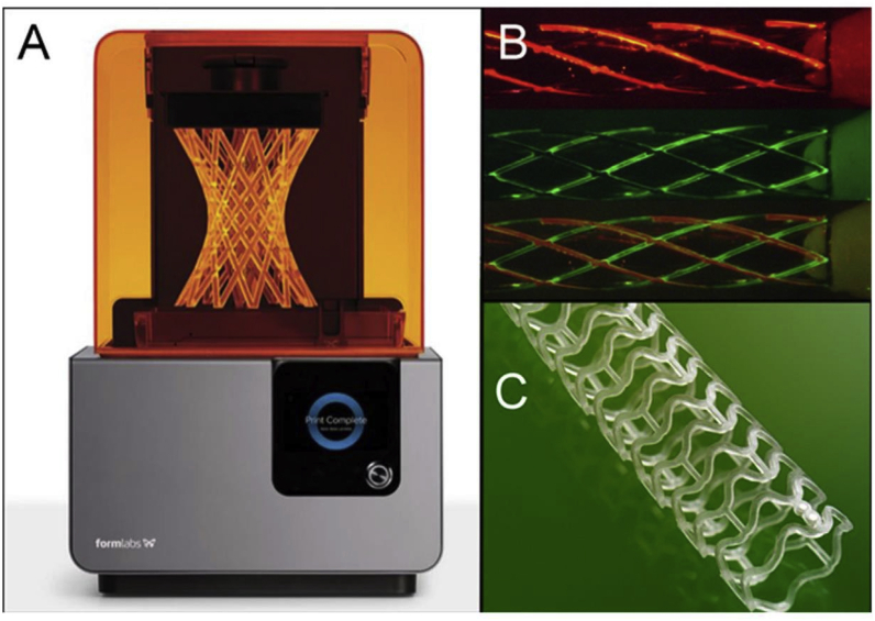
(A) Liquid-based 3D printers using laser to harden resin to create 3D structure (Courtesy: Formlab© 2015, Somerville, MA). (B) Prototype stents can be printed using different materials including metal (indicated by different colors). (C) A PLA bioabsorbable 3DP stent that is commercially available.
Transcatheter aortic procedure performed within an aortic stenosis model.
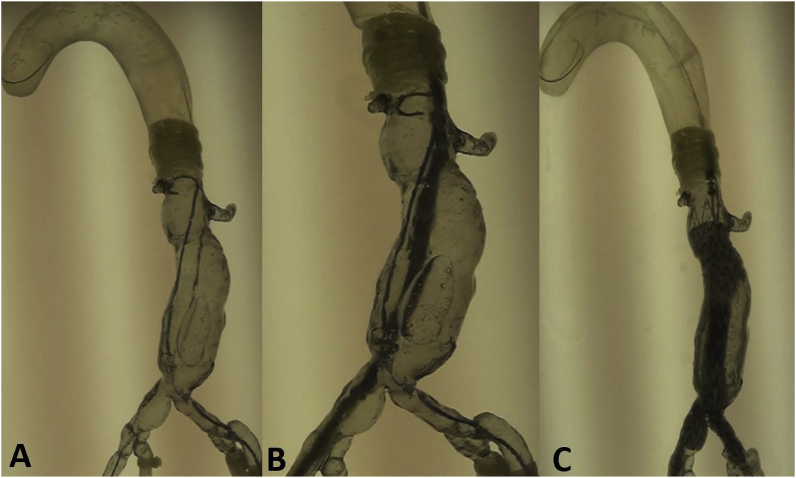
EVAR steps simulated in a 3D model. (A) Guidewire and catheter in position. (B) Main body of the stent graft in position. (C) Stent graft deployed.
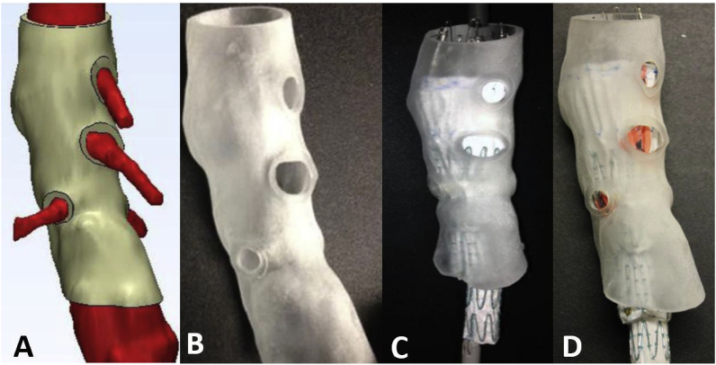
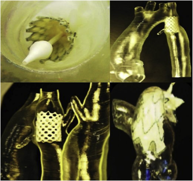
Process of determining fenestration locations on endograft. (A) Fenestration model created by computer software. (B) 3D printed fenestration model. (C) Stent graft is deployed inside the model. (D) Fenestration locations are marked onto the graft through the holes of the fenestration model.
Picture of 3D-printed AAA model (left) and its fluoroscopic image during the simulation (right).
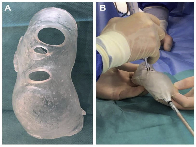
(A) 3DP model of the aortic arch. (B) Preparation of stent graft fenestration in the 3DP model.
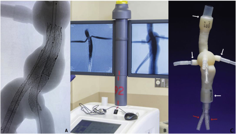
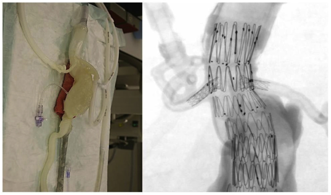
Fluoroscopy images (A) and EVAR simulation result (B) of 3D-printed AAA model with the attachment of plastic/silicone tubes connected to a fluid pump with water flow (C).
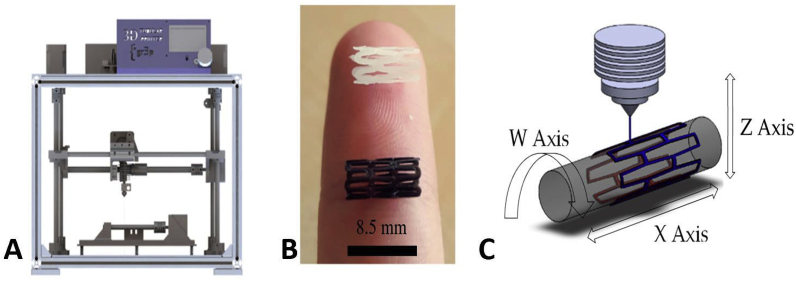
3D tubular printer (A) 3D-printed stents [PCL in white, PLA in black] (B), and printing methodology (C).
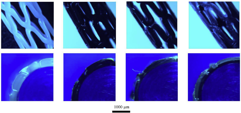
Optical microscope images of 3D-printed stents. The top row of images shows the general view, and the bottom row of images depicts the sectional view.
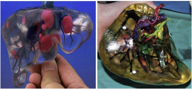
3D-printed liver cancer model. Different colors indicate blood vessels, biliary tract, and tumors.
Peripheral real-size vasculature from a CT angiogram and printed using red PLA.
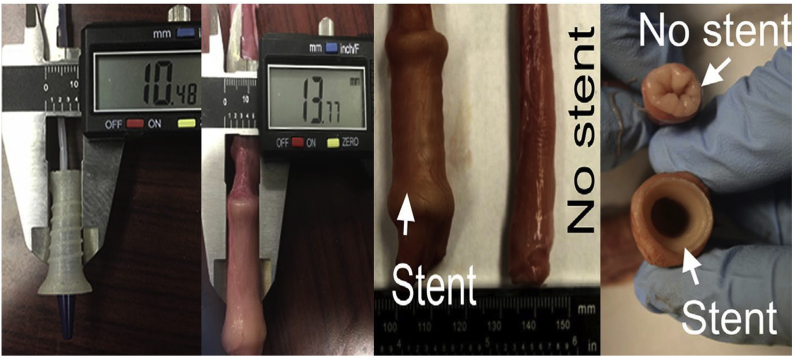
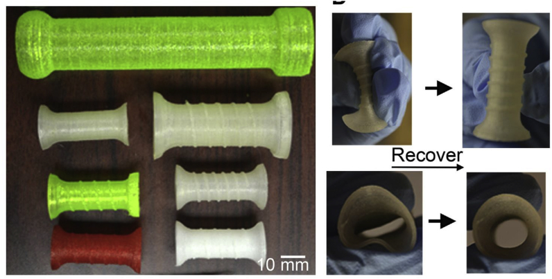
3DP personalized airway stent. (A) Images show the transition between the temporary state (for deployment) and the permanent shape (for performance). (B) Axial view of the process.
3DP esophagus stent and released state in the esophagus.
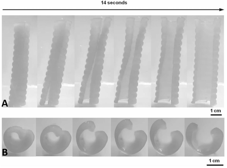
Different types of 3DP esophagus stents of different structures and materials (left). The stent was compressed and then recovered to the original shape (right).
(A) Upper gastrointestinal tract prototype STL file. (B). 3D-printed silicone moldings. (C) Assembled silicone moldings.
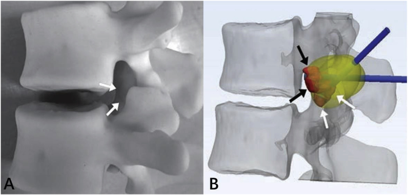
3D-printed model of L1 osteoblastoma; arrows indicate inferior palpable tumor lobule (A). (B) Advanced 3D visualization displays cryoprobe tracts (blue) used during the model simulation and theoretic ablation zones (yellow). Red indicates the tumor lobules. (For interpretation of the references to color in this figure legend, the reader is referred to the Web version of this article.)
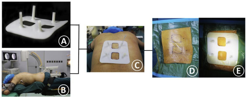
(A) 3D-printed guiding template for precise PVP. (B) Patient’s intraoperative position. (C) Matching the two location holes. (D) Marking the skin entry points and disinfect the surgery area. (E) Fixing the sterile guide template on the back skin.
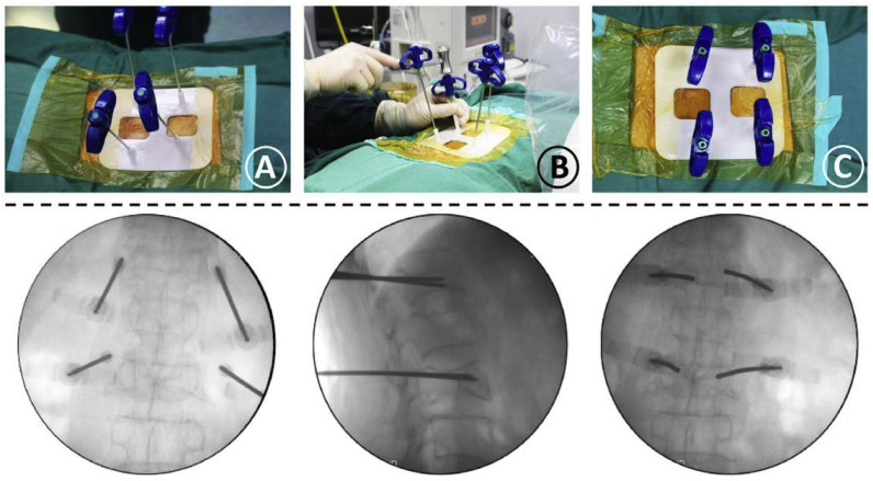
(A) Confirmation of the needles’ location. (B) The needles were gradually inserted into pedicles along the guiding cylinders. (C) The needles were completely inserted into the guiding cylinders of the template, and its final location was observed in the fluoroscopic images.
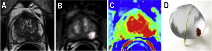
(A–C) Multiparametric MRI prostate transverse view. (D) 3D-printed model of a patient-specific prostate with tumor structure (red) and urethra (green). (For interpretation of the references to color in this figure legend, the reader is referred to the Web version of this article.)
Similar articles
-
Systematic review of three-dimensional printing for simulation training of interventional radiology trainees.3D Print Med. 2021 Apr 21;7(1):10. doi: 10.1186/s41205-021-00102-y. 3D Print Med. 2021. PMID: 33881672 Free PMC article.
-
Three-dimensional printing in cardiac surgery and interventional cardiology: a single-centre experience.Eur J Cardiothorac Surg. 2015 Jun;47(6):1044-52. doi: 10.1093/ejcts/ezu310. Epub 2014 Aug 26. Eur J Cardiothorac Surg. 2015. PMID: 25161184
-
Hybrid 3D printing: a game-changer in personalized cardiac medicine?Expert Rev Cardiovasc Ther. 2015 Dec;13(12):1281-4. doi: 10.1586/14779072.2015.1100076. Epub 2015 Oct 14. Expert Rev Cardiovasc Ther. 2015. PMID: 26465262
-
Three-dimensional printing in structural heart disease and intervention.Ann Transl Med. 2019 Oct;7(20):579. doi: 10.21037/atm.2019.09.73. Ann Transl Med. 2019. PMID: 31807560 Free PMC article. Review.
-
Clinical application of three-dimensional printing technology in craniofacial plastic surgery.Arch Plast Surg. 2015 May;42(3):267-77. doi: 10.5999/aps.2015.42.3.267. Epub 2015 May 14. Arch Plast Surg. 2015. PMID: 26015880 Free PMC article. Review.
Cited by
-
Erratum: An erratum on "Application of three-dimensional printing in interventional medicine" [J Interv Med (February 2020) 1-16].J Interv Med. 2020 Sep 19;3(3):156. doi: 10.1016/j.jimed.2020.06.001. eCollection 2020 Sep. J Interv Med. 2020. PMID: 34826305 Free PMC article.
-
Emerging Trends in Microfluidic Biomaterials: From Functional Design to Applications.J Funct Biomater. 2025 May 8;16(5):166. doi: 10.3390/jfb16050166. J Funct Biomater. 2025. PMID: 40422832 Free PMC article. Review.
-
3D Printing Techniques and Their Applications to Organ-on-a-Chip Platforms: A Systematic Review.Sensors (Basel). 2021 May 10;21(9):3304. doi: 10.3390/s21093304. Sensors (Basel). 2021. PMID: 34068811 Free PMC article.
-
Feasibility and practicality of a novel teaching aid for microvascular anastomosis simulation training in neurosurgery generated by 3D printing.Front Surg. 2025 Apr 30;12:1546573. doi: 10.3389/fsurg.2025.1546573. eCollection 2025. Front Surg. 2025. PMID: 40370766 Free PMC article.
-
Research on the application of collaborative teaching between ultrasonographers and vascular surgeons in the training of vascular surgery residents.Updates Surg. 2025 Aug;77(4):1233-1238. doi: 10.1007/s13304-025-02107-2. Epub 2025 Feb 1. Updates Surg. 2025. PMID: 39893346
References
-
- Becker H. Hype, hope and hubris: the quest for the killer application in microfluidics. Lab Chip. 2009;9:2119–2122. - PubMed
-
- Liaw C.Y., Guvendiren M. Current and emerging applications of 3D printing in medicine. Biofabrication. 2017;9 - PubMed
-
- Waheed S., Cabot J.M., Macdonald N.P. 3D printed microfluidic devices: enablers and barriers. Lab Chip. 2016;16:1993–2013. - PubMed
-
- Krujatz F., Lode A., Seidel J. Additive biotech-chances, challenges, and recent applications of additive manufacturing technologies in biotechnology. N Biotech. 2017;39:222–231. - PubMed
LinkOut - more resources
Full Text Sources