Review
. 2022 Sep;16(3):857-864.
doi: 10.1007/s12105-021-01394-6. Epub 2021 Nov 22.

Intrasalivary Thymic Carcinoma: A Case Report and Literature Review

Affiliations
Review

Intrasalivary Thymic Carcinoma: A Case Report and Literature Review

Michał Kunc et al. Head Neck Pathol. 2022 Sep.

Abstract

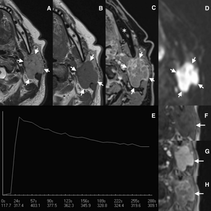
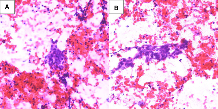
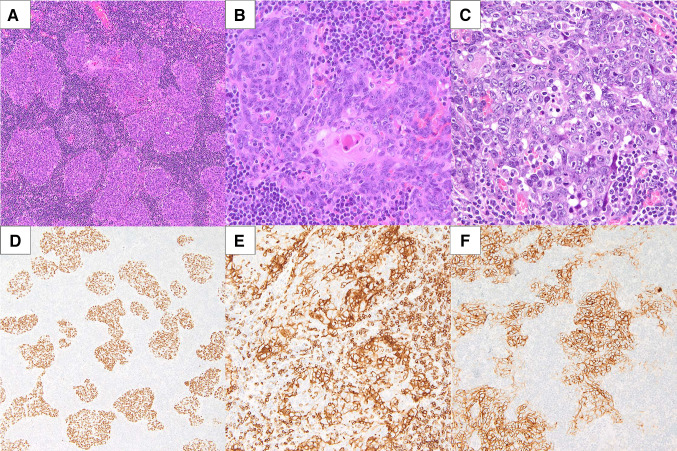
Ectopic thymic carcinomas are rarely diagnosed in the thyroid gland, let alone in extrathyroid tissues. In the currently available literature, only five cases of extrathyroidal malignancies with thymic differentiation have been reported as arising in the major salivary glands. A 69-year-old female presented with a slow-growing palpable mass in the left parotid gland. Fine needle aspiration biopsy suggested metastatic cancer, whereas core needle biopsy revealed high-grade squamous cell carcinoma. The patient underwent left radical parotidectomy with selective ipsilateral lymph node dissection and subsequent radiation therapy. The surgical specimen was taken for histopathological examination. Microscopically, the tumor resembled thymic carcinoma. It was composed of large nests of squamoid cells with smooth contours, focally with a syncytial growth pattern, and accompanied by abundant lymphocytes with reactive lymphoid follicles. This appearance resembled a micronodular thymic carcinoma with lymphoid hyperplasia. Moreover, the tumor displayed expression of squamous markers (p40 and p63) and markers of thymic carcinoma (CD5 and CD117). Therefore, the final diagnosis of intrasalivary thymic carcinoma was rendered. The molecular analysis including next-generation sequencing demonstrated no variants of the strong, potential, or unknown clinical significance. The patient remains disease-free at 1-year follow-up. In the current case, we comprehensively present a clinical, microscopic, molecular, and radiological picture of CD5-positive squamous cell carcinoma of the parotid. We postulate that similar cases should be designated as intrasalivary thymic carcinoma analogically to similar thyroid tumors. Our case and the limited literature data indicate they should be distinguished from conventional squamous cell carcinoma of major salivary glands due to their rather favorable prognosis.

Keywords: CASTLE; CD117; CD5; Immunohistochemistry; Parotid gland; Salivary gland; Squamous cell carcinoma; Thymic carcinoma.


Conflict of interest statement

The authors declare that they have no conflict of interest.

Figures

Fig. 1
Fig. 1
Magnetic resonance imaging in the axial plane of the left parotid region. In the turbo spin-echo sequence in T2-weighted image (a), the tumor (marked by the arrows) has intermediate signal intensity, observed in hypercellular lesions. In the turbo spin-echo sequence in T1-weighted image (b), the signal intensity is low. In the delayed contrast-enhanced gradient-echo T1-weighted image (c), the lesion shows heterogeneous and circumferential enhancement. In diffusion-weighted image (d) a marked diffusion restriction is observed (0.60–0.67 × 10−3 mm2/s), also typical for hypercellular lesions. The enhancement curve (e) shows the lesion’s signal intensity change after contrast agent administration on a timeline (expressed in seconds); early enhancement is visible with wash-out of the contrast agent (enhancement curve type B with wash-out ratio of 38%, typically seen in Warthin tumor). On pictures f to h, the subsequent phases of the dynamic examination are presented: pre-contrast image (f), early, strong, almost homogeneous enhancement of the lesion (g), and the last phase of the dynamic examination (h). Asterisk marks the mandible
Fig. 2
Fig. 2
Haematoxylin and eosin stained fine needle aspiration smears from the parotid tumor. Groups of pleomorphic, epithelioid cancer cells (A, B) in the lymphocytic background
Fig. 3
Fig. 3
Microscopic appearance of the tumor resembling micronodular thymic squamous cell carcinoma with lymphoid hyperplasia–cohesive growth, nests of cancer cells with smooth contours, prominent nucleoli, areas of medullary differentiation, and abundant lymphoid infiltrates with reactive lymphoid follicles formation (A). On higher magnification a structure reminiscent of Hassall’s corpuscle is visible (B). In some areas cancer cells demonstrate prominent nuclear pleomorphism, conspicuous nucleoli and multiple mitotic figures (C). Positive p63 staining in cancer cells emphasizes the micronodular architecture (D). CD5 is expressed by both cancer cells and some reactive lymphocytes (E), whereas CD117 is expressed exclusively by squamous cell carcinoma (F)

References

    1. Bai Y, Kakudo K, Jung CK. Updates in the pathologic classification of thyroid neoplasms: a review of the World Health Organization Classification. Endocrinol Metab. 2020;35:696–715. doi: 10.3803/EnM.2020.807. - DOI - PMC - PubMed
    1. Reimann JDR, Dorfman DM, Nosé V. Carcinoma showing thymus-like differentiation of the thyroid (CASTLE): a comparative study. Am J Surg Pathol. 2006;30:994–1001. doi: 10.1097/00000478-200608000-00010. - DOI - PubMed
    1. Gordon J, Manley NR. Mechanisms of thymus organogenesis and morphogenesis. Development. 2011;138:3865–3878. doi: 10.1242/dev.059998. - DOI - PMC - PubMed
    1. Fukushima T, Suzuki S, Ohira T, Shimura H, Midorikawa S, Ohtsuru A, et al. Prevalence of ectopic intrathyroidal thymus in Japan: the Fukushima Health Management Survey. Thyroid. 2015;25:534–537. doi: 10.1089/thy.2014.0367. - DOI - PubMed
    1. Ito Y, Miyauchi A, Nakamura Y, Miya A, Kobayashi K, Kakudo K. Clinicopathologic significance of intrathyroidal epithelial thymoma/carcinoma showing thymus-like differentiation: a collaborative study with member Institutes of the Japanese Society of Thyroid Surgery. Am J Clin Pathol. 2007;127:230–236. doi: 10.1309/VM7E52B6U9Q729DQ. - DOI - PubMed