. 2021 Nov 22;16(11):e0254282.
doi: 10.1371/journal.pone.0254282. eCollection 2021.

Origin and evolutionary landscape of Nr2f transcription factors across Metazoa

Affiliations

Origin and evolutionary landscape of Nr2f transcription factors across Metazoa

Ugo Coppola et al. PLoS One. .

Abstract

Background: Nuclear Receptor Subfamily 2 Group F (Nr2f) orphan nuclear hormone transcription factors (TFs) are fundamental regulators of many developmental processes in invertebrates and vertebrates. Despite the importance of these TFs throughout metazoan development, previous work has not clearly outlined their evolutionary history.

Results: We integrated molecular phylogeny with comparisons of intron/exon structure, domain architecture, and syntenic conservation to define critical evolutionary events that distinguish the Nr2f gene family in Metazoa. Our data indicate that a single ancestral eumetazoan Nr2f gene predated six main Bilateria subfamilies, which include single Nr2f homologs, here referred to as Nr2f1/2/5/6, that are present in invertebrate protostomes and deuterostomes, Nr2f1/2 homologs in agnathans, and Nr2f1, Nr2f2, Nr2f5, and Nr2f6 orthologs that are found in gnathostomes. Four cnidarian Nr2f1/2/5/6 and three agnathan Nr2f1/2 members are each due to independent expansions, while the vertebrate Nr2f1/Nr2f2 and Nr2f5/Nr2f6 members each form paralogous groups that arose from the established series of whole-genome duplications (WGDs). Nr2f6 members are the most divergent Nr2f subfamily in gnathostomes. Interestingly, in contrast to the other gnathostome Nr2f subfamilies, Nr2f5 has been independently lost in numerous vertebrate lineages. Furthermore, our analysis shows there are differential expansions and losses of Nr2f genes in teleosts following their additional rounds of WGDs.

Conclusion: Overall, our analysis of Nr2f gene evolution helps to reveal the origins and previously unrecognized relationships of this ancient TF family, which may allow for greater insights into the conservation of Nr2f functions that shape Metazoan body plans.


Conflict of interest statement

The authors have declared that no competing interests exist.

Figures

Fig 1
Fig 1. Schematic of conserved domain architecture of Nr2f TFs.
A/B (N-terminal variable domain with transactivating AF-1 domain), C (DBD, which contains two Zinc finger (Znf) motifs), D (a linker domain), E (LBD plus transactivating AF-2 domain), and F (C-terminal).
Fig 2
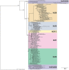
Fig 2. Evolutionary reconstruction of Nr2f TFs in metazoans.
Phylogenetic tree of Nr2f members demonstrate the existence of six protein classes: Nr2f1/2/5/6 (violet box), Nr2f1/2 (yellow box), Nr2f1 (pink box), Nr2f2 (green box), Nr2f5 (blue box), Nr2f6 (orange box). The placozoan Trichoplax adhaerens Nr2f was used as the outgroup. Values at the branches indicate replicates obtained using the aLRT method.
Fig 3
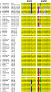
Fig 3. Zinc finger (Znf) motifs within the DBD of the Nr2f family.
Alignments of first (I) and second (II) Znfs found in Nr2f TFs. Yellow represents highly conserved amino acids throughout all species. White indicates amino acids that are not conserved. Turquoise and blue indicate amino acid changes that are conserved within Znf I of Nr2f5 and -6, respectively. The valine change found in some Nr2f6 LBDs is also found in the placozoan and cnidaria Nr2fs. Magenta and red indicate amino acid changes at the same residue that are conserved within Znf II of Nr2f5 and Nr2f6, respectively. A glycine residue is also found at the same position in some cnidaria and invertebrate Nr2fs. Green indicates a conserved change found in most Nr2f5 and Nr2f6 Znf IIs.
Fig 4
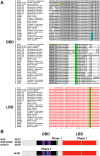
Fig 4. Intron code of the Nr2f family in metazoans.
(A) Protein alignment showing conservation of intron/exon structures within the DBDs (black) and LBDs (red) of Nr2f members. Znfs in the DBDs are underlined. Phase 0 introns—yellow, phase 1 introns—green, and phase 2 introns—turquoise. Asterisks indicate 100% amino acid conservation. Colons indicate high levels (>90%) amino acid conservation. Periods indicate moderate levels (50–89%) of amino acid conservation. (B) Schematization of intron/exon boundaries of Nr2f genes as they relate the Nr2f protein DBD and LBDs. Black box indicates DBD. Purple boxes represent the zinc-fingers motifs within the DBD. Red boxes indicate the LDB. Colored bars indicate the conserved Nr2f Phase 1 introns (green) and the Nr2f6-specific Phase 2 intron (turquoise).
Fig 5
Fig 5. Synteny analysis of Nr2f1/2/5/6 genes in cnidaria.
Schematic of the loci flanking Nr2f1/2/5/6 gene duplications in cnidaria N. vectensis, A. millepora, and H vulgaris. Only the one H. vulgaris gene presently has available genomic information. Arrows indicate transcription orientation.
Fig 6
Fig 6. Synteny analysis of vertebrate Nr2f1 and Nr2f2 genes.
Schematization of conserved genomic environments of gnathostome Nr2f1 and Nr2f2 genes (red rectangles) in selected species with relative chromosomes/scaffolds. Flanking orthologous genes are represented employing rectangles of the same color. Arrows indicate transcription orientation.
Fig 7
Fig 7. Synteny analysis of vertebrate Nr2f5 genes.
Schematization of conserved genomic environments of gnathostome Nr2f5 genes (red rectangles) in selected species with relative chromosomes/scaffolds. Flanking orthologous genes are represented using rectangles of the same color. Arrows indicate transcription orientation.
Fig 8
Fig 8. Synteny analysis of vertebrate Nr2f6 genes.
Schematization of conserved genomic environments of gnathostome Nr2f6 genes (red rectangles) in selected species with relative chromosomes/scaffolds. Flanking orthologous genes are represented using the same color code. Arrows indicate transcription orientation.
Fig 9
Fig 9. Synteny analysis of Nr2f genes in teleosts.
Comparison of Nr2f genome environments in selected teleosts (zebrafish, Asian arowana, Atlantic salmon) with relative chromosomes/scaffolds. Rectangles of the same color represent flanking orthologous genes. Arrows indicate transcription orientation.
Fig 10
Fig 10. Model summarizing the evolutionary events of the Nr2f family in Metazoa.
A single Nr2f of placozoans (white box) represents the ancestor of extant Nr2fs. There were duplicative events specific to cnidaria leading to the expansion of Nr2f1/2/5/6 (pink circle). Invertebrate protostomes and deuterostomes have predominantly retained a single Nr2f1/2/5/6 homolog. There were duplicative events specific to agnathans leading to an expansion of Nr2f1/2 genes (orange circle). WGDs within vertebrates (green circles) generated the four Nr2fs found in vertebrates, with Nr2f1/Nr2f2 being paralogous and Nr2f5/Nr2f6 being paralogous. Nr2f5 has been independently lost in multiple vertebrate groups (red circle). It is lost in some cartilaginous fish and turtles (reptilian amniotes), and is absent in avian and mammalian amniotes. Teleosts have additional Nr2f Ohnologs due to TSGDs (blue circles) and SSGDs (yellow circles).

References

    1. Laudet V. Evolution of the nuclear receptor superfamily: early diversification from an ancestral orphan receptor. J Mol Endocrinol. 1997;19: 207–226. doi: 10.1677/jme.0.0190207 - DOI - PubMed
    1. Escriva H, Safi R, Hänni C, Langlois MC, Saumitou-Laprade P, Stehelin D, et al.. Ligand binding was acquired during evolution of nuclear receptors. Proc Natl Acad Sci U S A. 1997;94: 6803–6808. doi: 10.1073/pnas.94.13.6803 - DOI - PMC - PubMed
    1. Escriva H, Bertrand S, Laudet V. The evolution of the nuclear receptor superfamily. Essays in Biochemistry. 2004. pp. 11–26. doi: 10.1042/bse0400011 - DOI - PubMed
    1. Mangelsdorf DJ, Evans RM. The RXR heterodimers and orphan receptors. Cell. 1995. pp. 841–850. doi: 10.1016/0092-8674(95)90200-7 - DOI - PubMed
    1. Pastorcic M, Wang H, Elbrecht A, Tsai SY, Tsai MJ, O’Malley BW. Control of transcription initiation in vitro requires binding of a transcription factor to the distal promoter of the ovalbumin gene. Molecular and Cellular Biology. 1986. pp. 2784–2791. doi: 10.1128/mcb.6.8.2784-2791.1986 - DOI - PMC - PubMed

Publication types

Substances