. 2021 Dec;27(12):3092-3102.
doi: 10.3201/eid2712.211088.

Mammarenaviruses of Rodents, South Africa and Zimbabwe

Mammarenaviruses of Rodents, South Africa and Zimbabwe

Antoinette A Grobbelaar et al. Emerg Infect Dis. 2021 Dec.

Abstract

We conducted a survey for group-specific indirect immunofluorescence antibody to mammarenaviruses by using Lassa fever and Mopeia virus antigens on serum specimens of 5,363 rodents of 33 species collected in South Africa and Zimbabwe during 1964-1994. Rodents were collected for unrelated purposes or for this study and stored at -70°C. We found antibody to be widely distributed in the 2 countries; antibody was detected in serum specimens of 1.2%-31.8% of 14 species of myomorph rodents, whereas 19 mammarenavirus isolates were obtained from serum specimens and viscera of 4 seropositive species. Phylogenetic analysis on the basis of partial nucleoprotein sequences indicates that 14 isolates from Mastomys natalensis, the Natal multimammate mouse, were Mopeia virus, whereas Merino Walk virus was characterized as a novel virus in a separate study. The remaining 4 isolates from 3 rodent species potentially constitute novel viruses pending full characterization.

Keywords: Mastomys natalensis; Mopeia virus; Natal multimammate mouse; South Africa; Zimbabwe; mammarenavirus; rodents; viruses.


Figures

Figure 1
Figure 1
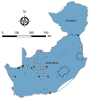
Locations where samples from 5,363 rodents of 33 species were collected and tested for evidence of infection with mammarenaviruses, South Africa and Zimbabwe. White circles indicate sites where no evidence of infection was found; black circles indicate sites where antibody to mammarenaviruses was detected by indirect immunofluorescence.
Figure 2
Figure 2
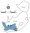
Locations where samples were collected from Mastomys spp. rodents, South Africa and Zimbabwe. White squares indicate sites where no antibody to mammarenaviruses was found in M. coucha mouse serum specimens; black squares,where antibody was detected in M. coucha mouse serum specimens; white circles, where no antibody to mammarenaviruses was found in M. natalensis mouse serum specimens; black circles, where antibody was detected in M. natalensis mouse serum specimens; black triangles, where Mopeia virus was isolated from M. natalensis mouse samples during this study; black diamonds, where Mopeia virus was isolated from M. natalensis mouse samples during previous studies, including the original isolations in Mozambique (9,10). Shading indicates distribution ranges for M. coucha and M. natalensis mice. Adapted from Chimimba and Bennett (15).
Figure 3
Figure 3
Locations where samples were collected from Micaelamys namaquensis rodents, South Africa and Zimbabwe. White circles indicate sites where no antibody to mammarenaviruses was found in M. namaquensis rat serum specimens; black circles, where antibody was detected in M. namaquensis rat serum specimens; black triangle, where a mammarenavirus isolate was obtained from an M. namaquensis rat sample. Shading indicates distribution range of M. namaquensis rats. Adapted from Chimimba and Bennett (15).
Figure 4
Figure 4
Locations where samples were collected from Otomys unisulcatus rodents, South Africa and Zimbabwe. White circles indicate sites where no antibody to mammarenaviruses was found in O. unisulcatus rat serum specimens; black circles, where antibody was detected in O. unisulcatus rat serum specimens; black triangle, where Omdraaivlei mammarenavirus isolates were obtained from O. unisulcatus rat samples; black diamond, where Merino Walk virus was isolated from O. unisulcatus rat. Shading indicates distribution range of O. unisulcatus rats. Adapted from Chimimba and Bennett (15).
Figure 5
Figure 5
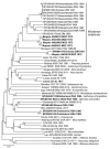
Phylogenetic relationships of 48 arenavirus isolates based on neighbor-joining analysis applying a Jukes-Cantor model of partial nucleoprotein sequences (≈912 nt), together with known host relationships and collection dates. Values at nodes indicate the level (%) of bootstrap support from 1,000 replicates. Scale bar indicates base substitutions per site. Bold indicates sequences determined in this study. ANGL, Angola; AR, Argentina; AU, Australia; CAM, Cameroon; CAR, Central Africa Republic; CO, Colombia; CIV, Côte d'Ivoire; ETH, Ethiopia; GHA, Ghana; MOZ, Mozambique; NAMB, Namibia; NIG, Nigeria; RSA, Republic of South Africa; SL, Sierra Leone; TAN, Tanzania; TR, Trinidad; USA, United States; ZAM, Zambia; ZIM, Zimbabwe.
Figure 6
Figure 6
Phylogenetic relationships of 8 rodents from which mammarenavirus isolates were obtained in this study (bold) and reference taxonomic voucher sequences from GenBank. Tree was constructed based on neighbor-joining analysis of a 136-bp cytochrome b barcode sequence. Values at nodes indicate the level of bootstrap support from 1,000 replicates. Scale bar indicates base substitutions per site. GenBank accession numbers, rodent reference number, and country of collection are indicated. RSA, Republic of South Africa; ZIM, Zimbabwe.

References

    1. Swanepoel R. Viral haemorrhagic fevers in South Africa: history and national strategy. S Afr J Sci. 1987;83:80–8.
    1. Swanepoel R, Shepherd AJ, Leman PA, Shepherd SP, McGillivray GM, Erasmus MJ, et al. Epidemiologic and clinical features of Crimean-Congo hemorrhagic fever in southern Africa. Am J Trop Med Hyg. 1987;36:120–32. 10.4269/ajtmh.1987.36.120 - DOI - PubMed
    1. Shepherd AJ, Swanepoel R, Shepherd SP, McGillivray GM, Searle LA. Antibody to Crimean-Congo hemorrhagic fever virus in wild mammals from southern Africa. Am J Trop Med Hyg. 1987;36:133–42. 10.4269/ajtmh.1987.36.133 - DOI - PubMed
    1. Johnson KM, Elliott LH, Heymann DL. Preparation of polyvalent viral immunofluorescent intracellular antigens and use in human serosurveys. J Clin Microbiol. 1981;14:527–9. 10.1128/jcm.14.5.527-529.1981 - DOI - PMC - PubMed
    1. Ksiazek TG, West CP, Rollin PE, Jahrling PB, Peters CJ. ELISA for the detection of antibodies to Ebola viruses. J Infect Dis. 1999;179(Suppl 1):S192–8. 10.1086/514313 - DOI - PubMed

LinkOut - more resources