. 2022 Jun;142(6):1597-1606.e9.
doi: 10.1016/j.jid.2021.10.025. Epub 2021 Nov 20.

Skin Fibrosis and Recovery Is Dependent on Wnt Activation via DPP4

Affiliations

Skin Fibrosis and Recovery Is Dependent on Wnt Activation via DPP4

Anna R Jussila et al. J Invest Dermatol. 2022 Jun.

Abstract

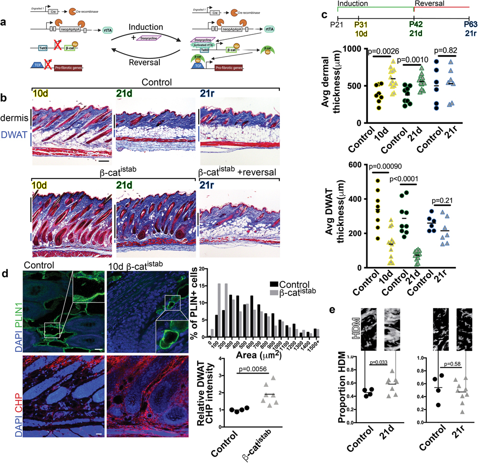
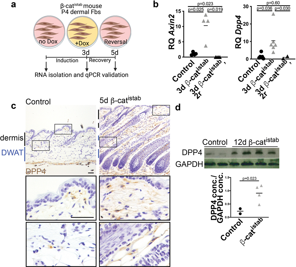
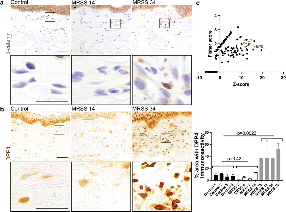
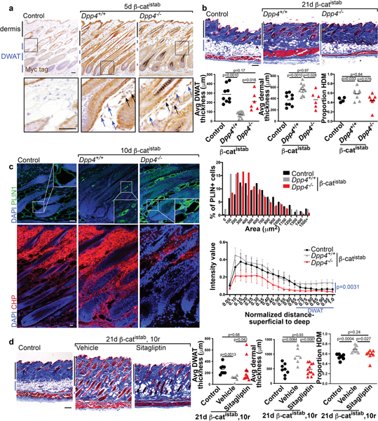
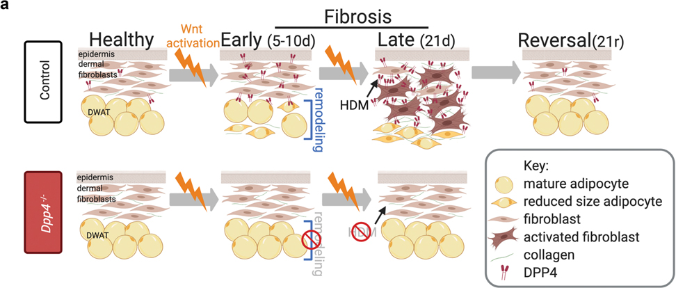
Fibrosis is the life-threatening, excessive accumulation of the extracellular matrix and is sometimes associated with a loss of lipid-filled cells in the skin and other organs. Understanding the mechanisms of fibrosis and associated lipodystrophy and their reversal may reveal new targets for therapeutic intervention. In vivo genetic models are needed to identify key targets that induce recovery from established fibrosis. Wnt signaling is activated in animal and human fibrotic diseases across organs. Here, we developed a genetically inducible and reversible Wnt activation model and showed that it is sufficient to cause fibrotic dermal remodeling, including extracellular matrix expansion and shrinking of dermal adipocytes. Upon withdrawal from Wnt activation, Wnt-induced fibrotic remodeling was reversed in mouse skin-fully restoring skin architecture. Next, we demonstrated CD26/ DPP4 is a Wnt/β-catenin-responsive gene and a functional mediator of fibrotic transformation. We provide genetic evidence that the Wnt/DPP4 axis is required to drive fibrotic dermal remodeling and is associated with human skin fibrosis severity. Remarkably, DPP4 inhibitors can be repurposed to accelerate recovery from established Wnt-induced fibrosis. Collectively, this study identifies Wnt/DPP4 axis as a key driver of extracellular matrix homeostasis and dermal fat loss, providing therapeutic avenues to manipulate the onset and reversal of tissue fibrosis.


Conflict of interest statement

Conflict of interest: RL has received consulting fees from Bristol Myers Squibb, Boehringer Ingelheim, Formation, Sanofi, Boehringer-Mannheim, Merck, Genentech/Roche, Certa, Pfizer, Magenta, Biogen and Formation, and grant support from Biogen, Corbus, Formation, Moderna, Regeneron, Pfizer, Kiniksa, Astra Zeneca, Kyowa, Kirin, and Genentech/Roche. The other authors state no conflict of interest.

Figures

Figure 1:
Figure 1:. β-catistab leads to inducible and reversible dermal remodeling in mice.
a, Transgenes in doxycycline-inducible/reversible β-catenin (β-catistab) dermal fibrosis model. b, Fibrosis progression in Masson’s trichrome stained control (top) and β-catistab (bottom) dermis; black and blue bars indicate dermal and DWAT thickness; scale bar=200μm. c, Quantification of average dorsal dermal and DWAT thickness/mouse (n=6–11). d, Indirect immunofluorescence of PLIN1 (green) and CHP stain (red) in control and 10d β-catistab DWAT; quantification of area of PLIN1+ vesicles, 50/mouse (n=9) and relative corrected fluorescence CHP intensity in DWAT; scale bar=25μm. e, Quantification of avg collagen high density matrix (HDM)/40x field/mouse with HDM mask (n=4–9).
Figure 2:
Figure 2:. DPP4 expression is Wnt responsive and elevated in β-catistab dermal fibrosis model.
a, Schematic of in vitro validation. b, Dpp4 and Wnt target gene, Axin2, expression by qRT-PCR relative to housekeeping gene, Hprt, in control, β-catistab, and reversal following β-catistab dermal cells. c, DPP4 immunohistochemical staining on control and 5d β-catistab mouse skin with high magnification panels; scale bar=100μm. d, DPP4 protein quantity relative to GAPDH protein quantity in whole skin by western blot.
Figure 3:
Figure 3:. DPP4 and β-catenin expression are elevated in high MRSS-scored human skin.
a, IHC with α-β-catenin antibody from left to right: Control, low MRSS scored (14) human forearm biopsy sample, high MRSS scored (34) human forearm biopsy sample; scale bar= 100μm in low mag, 50μm in high mag. b, IHC with α-DPP4 antibody on same samples as in a; accompanying quantification of 1–3 fields per control and scored SSc human forearm skin sample. c, Transcription factor binding site overrepresentation analyisis using oPPOSUM showing that promoter region of DPP4 is enriched for predicted binding sites for Wnt signaling TFs, including TCF7l2 and LEF1.
Figure 4:
Figure 4:. DPP4 mediates dermal remodeling in β-catistab dermal fibrosis model.
a, Myc-tag IHC in control, Dpp4+/+ 5d β-catistab, and Dpp4−/− 5d β-catistab dorsal dermis; blue arrows indicate adipocytes with high signal, black arrows indicate fibroblasts with high signal; scale bar=100μm. b, Masson’s trichrome stained dorsal skin of control, Dpp4+/+ 21d β-catistab, and Dpp4−/− 21d β-catistab; quantification of dermal and DWAT thickness/mouse (n=6–10) and proportion of collagen high density matrix (n=4–8); scale bar=200μm. c, Indirect immunofluorescence staining for PLIN1 (green) and CHP staining (red) in mouse skin from control, Dpp4+/+ 10d β-catistab, and Dpp4−/− 10d β-catistab mice with histogram of PLIN1+ vesicle size (n=6–9) and DWAT CHP intensity heatmap by skin depth with standard error bars; significance between DWAT region intensity values only; scale bar=25μm. d, Masson’s trichrome stained mouse skin from control, 21d β-catistab and 10d reversal with vehicle or sitagliptin treatment; scale bar=200μm; quantification of dermal, DWAT thickness, and HDM/mouse(n=6–11).
Figure 5:
Figure 5:. Summary schematic.
a, By 10d of β-catistab, adipocytes undergo DWAT remodeling ultimately leading to DWAT loss and collagen accumulation by 21d. DPP4 is Wnt-responsive and increased within 5d of β-catistab, prior to DWAT remodeling. Dpp4−/− mice have preservation of DWAT and reduced DWAT remodeling despite 10d β-catistab resulting in reduced dermal thickening, less high density matrix (HDM), and DWAT preservation after 21d of β-catistab.

References

    1. Agha El, E, Moiseenko A, Kheirollahi V, De Langhe S, Crnkovic S, Kwapiszewska G, et al., (2017). Two-Way Conversion between Lipogenic and Myogenic Fibroblastic Phenotypes Marks the Progression and Resolution of Lung Fibrosis. Cell Stem Cell 20: 261–3. - PMC - PubMed
    1. Akhmetshina A, Palumbo K, Dees C, Bergmann C, Venalis P, Zerr P, et al., (2012). Activation of canonical Wnt signalling is required for TGF-β-mediated fibrosis. Nature Communications 3: 735–12. - PMC - PubMed
    1. Alexander CM, Kasza I, Yen E, Reeder S, Hernando D, Gallo R, et al., (2015). Dermal white adipose tissue: a new component of the thermogenic response. J Lipid Res 56: 2061–9. - PMC - PubMed
    1. Atit R, Sgaier SK, Mohamed OA, Taketo M, Dufort D, Joyner A, et al., (2006). Beta-catenin activation is necessary and sufficient to specify the dorsal dermal fate in the mouse. Developmental Biology 296: 164–76. - PubMed
    1. Baticic Pucar L, Pernjak Pugel E, Detel D, Varlejen J (2017). Involvement of DPP IV/CD26 in cutaneous wound healing process in mice. Wound Repair Regen 25: 25–40. - PubMed

Publication types