

Spatiotemporal control of myofibroblast activation in acoustically-responsive scaffolds via ultrasound-induced matrix stiffening
- PMID: 34808418
- PMCID: PMC8738148
- DOI: 10.1016/j.actbio.2021.11.020
Spatiotemporal control of myofibroblast activation in acoustically-responsive scaffolds via ultrasound-induced matrix stiffening
Abstract
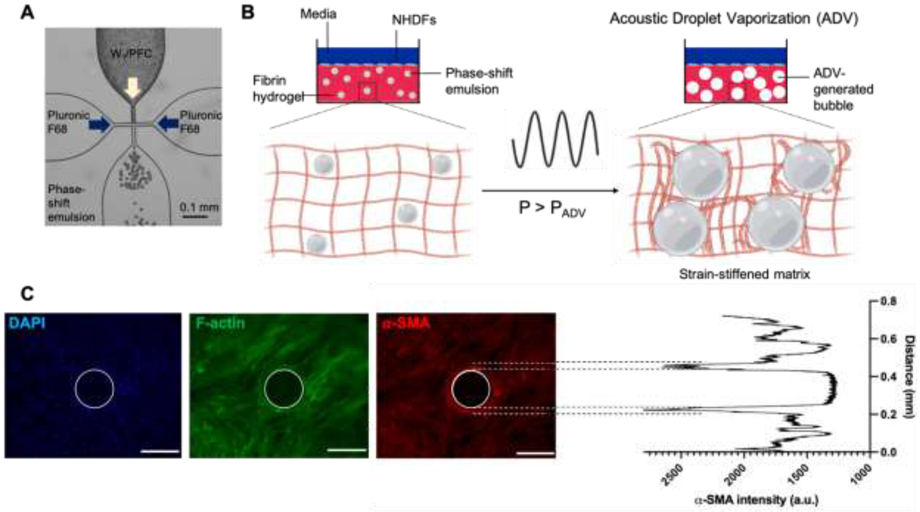
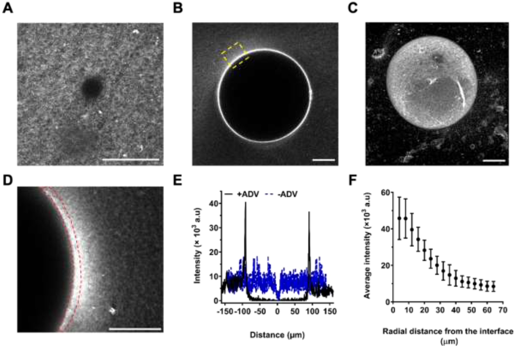
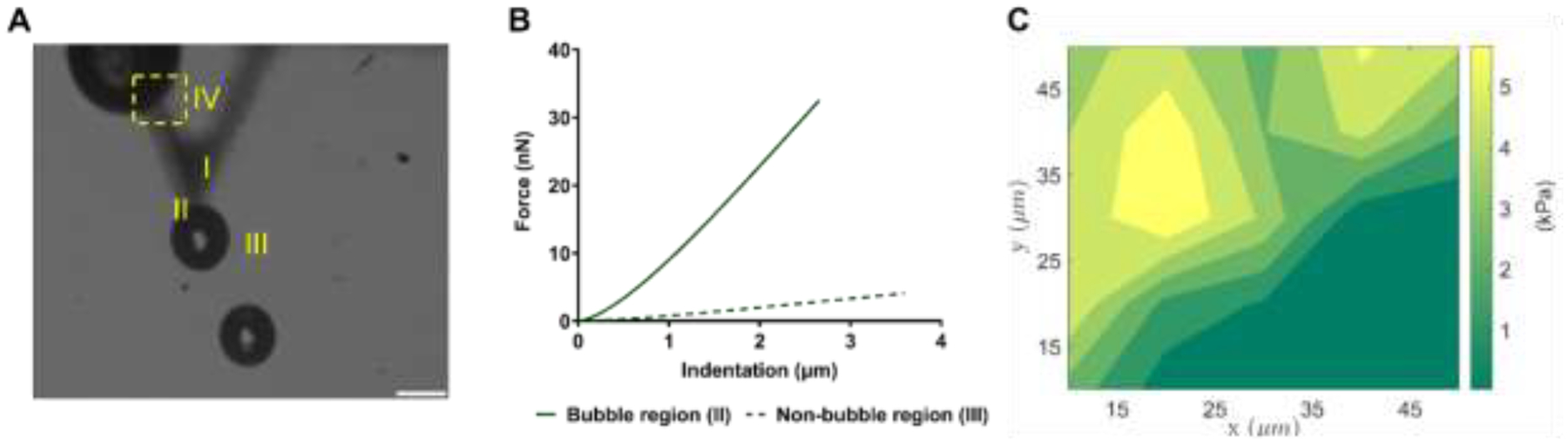
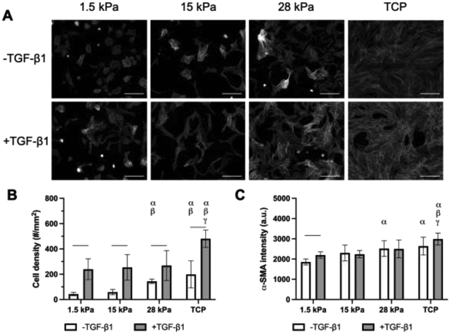
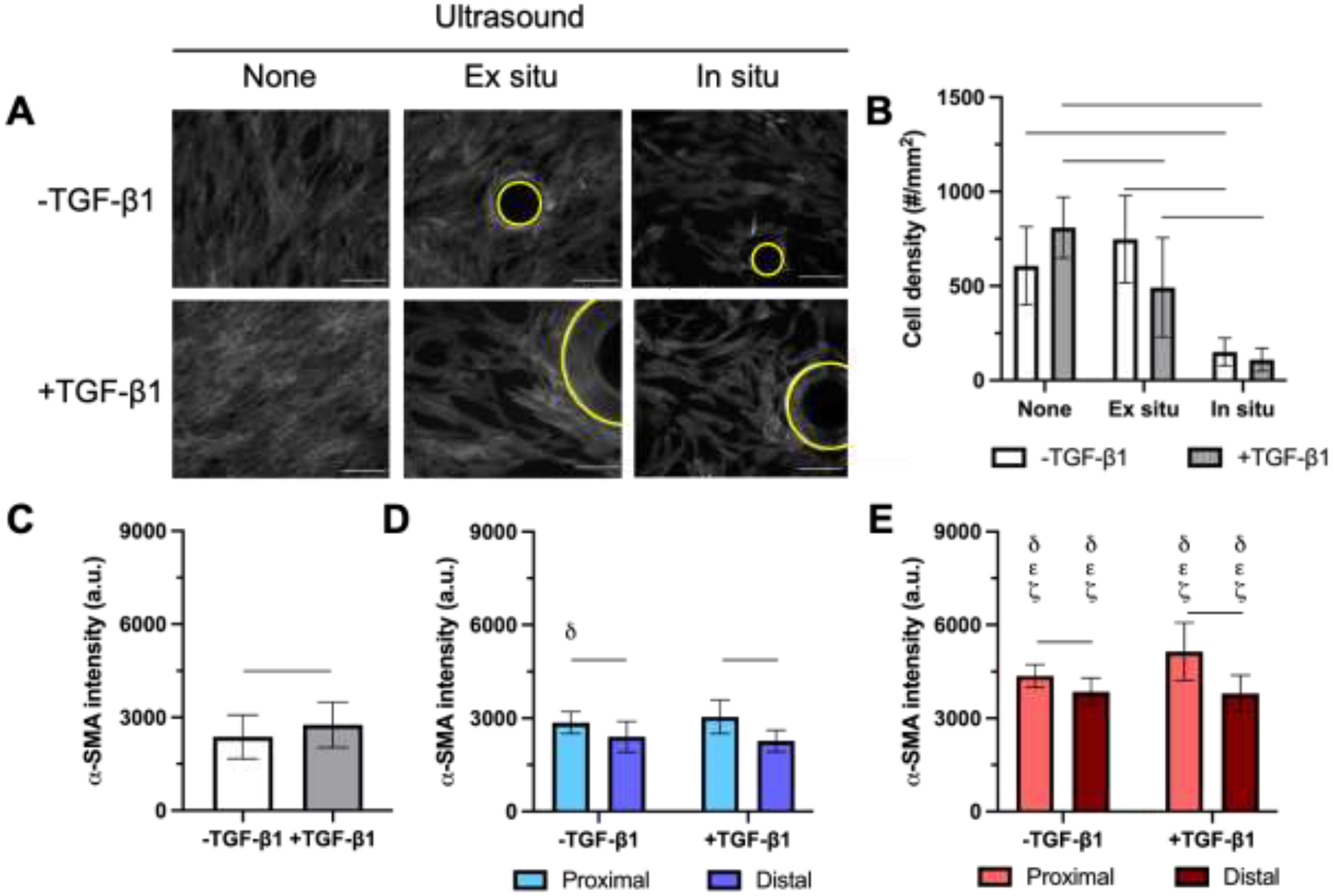
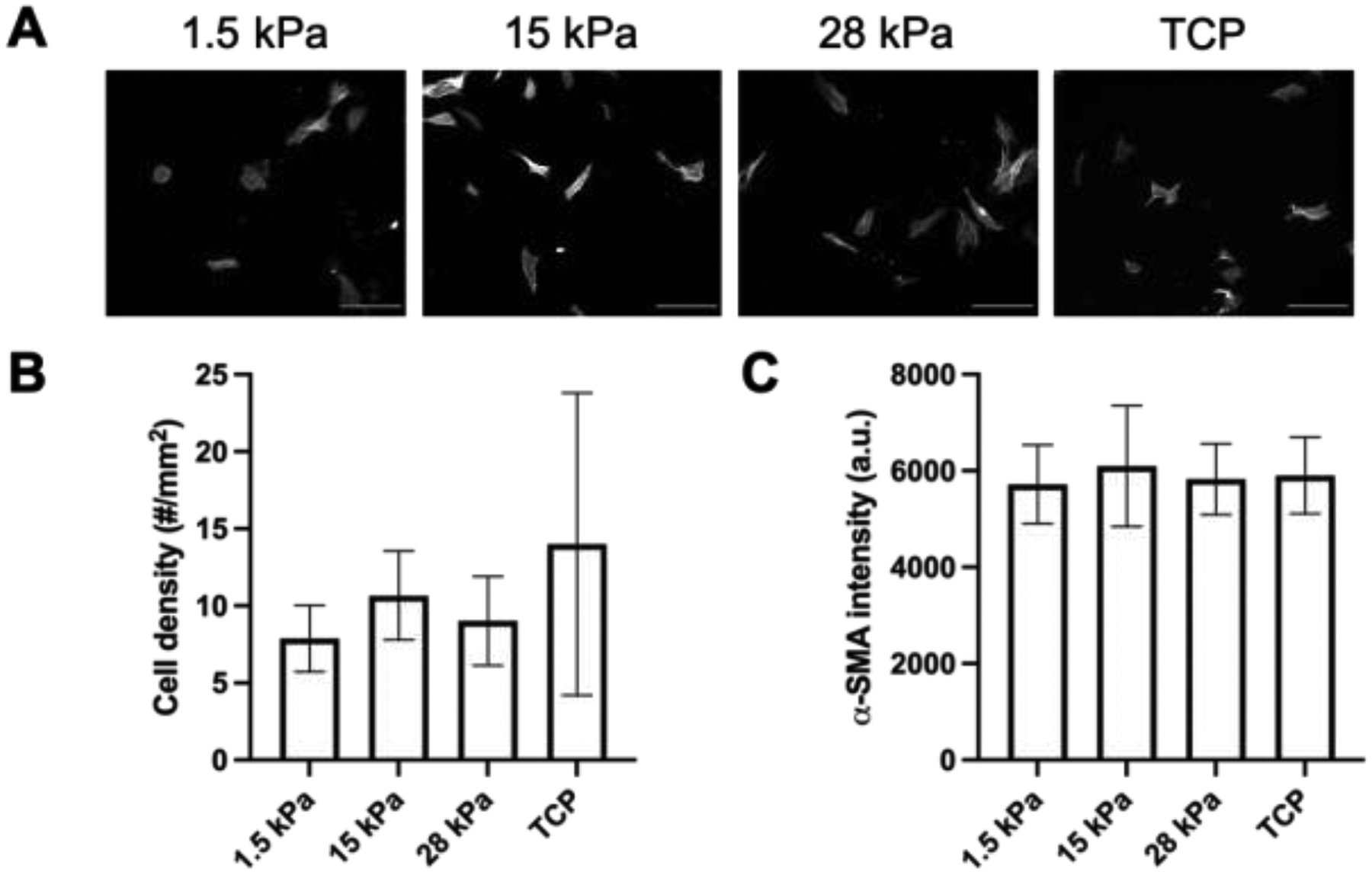
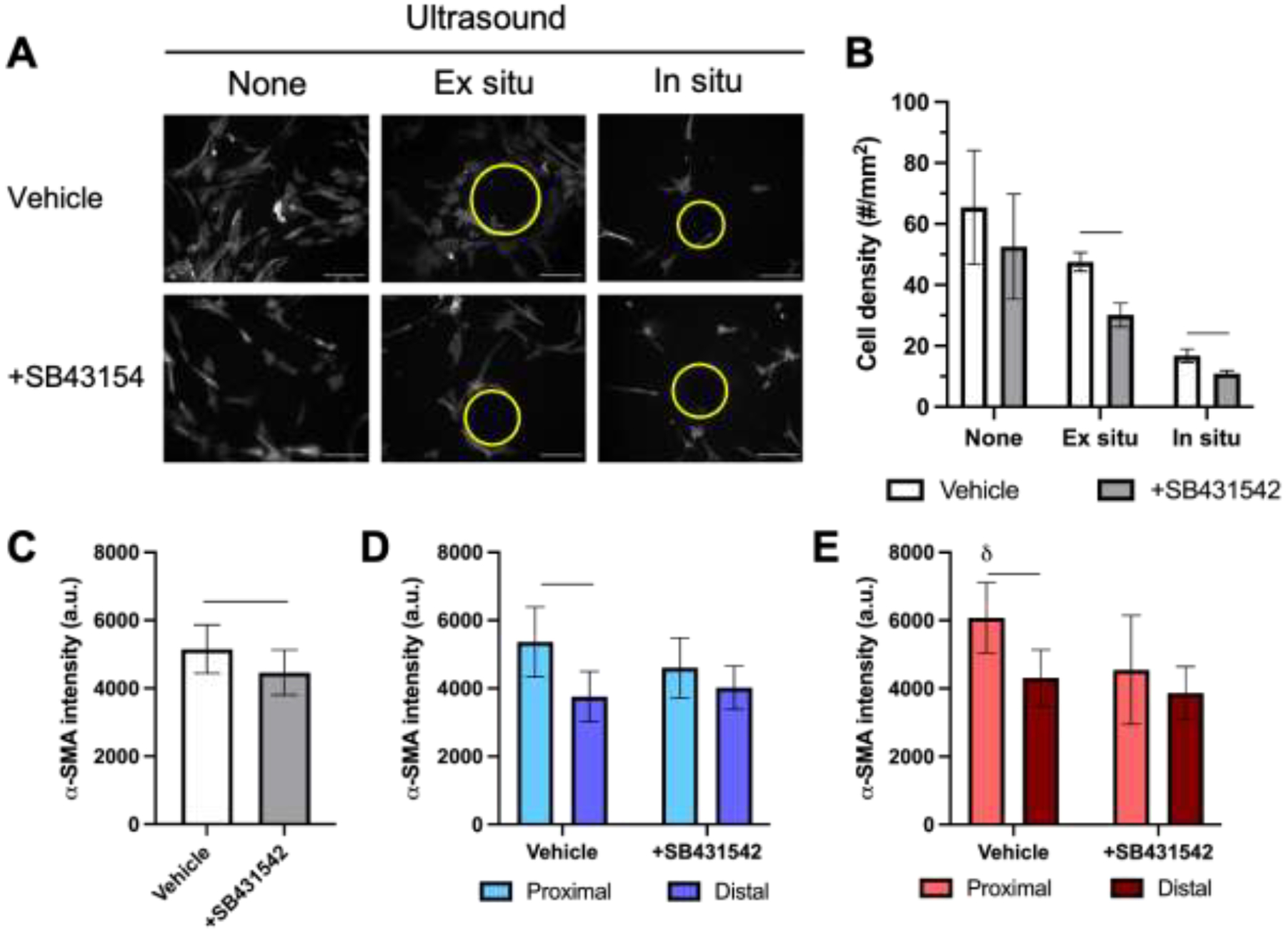
Hydrogels are often used to study the impact of biomechanical and topographical cues on cell behavior. Conventional hydrogels are designed a priori, with characteristics that cannot be dynamically changed in an externally controlled, user-defined manner. We developed a composite hydrogel, termed an acoustically-responsive scaffold (ARS), that enables non-invasive, spatiotemporally controlled modulation of mechanical and morphological properties using focused ultrasound. An ARS consists of a phase-shift emulsion distributed in a fibrin matrix. Ultrasound non-thermally vaporizes the emulsion into bubbles, which induces localized, radial compaction and stiffening of the fibrin matrix. In this in vitro study, we investigate how this mechanism can control the differentiation of fibroblasts into myofibroblasts, a transition correlated with substrate stiffness on 2D substrates. Matrix compaction and stiffening was shown to be highly localized using confocal and atomic force microscopies, respectively. Myofibroblast phenotype, evaluated by α-smooth muscle actin (α-SMA) immunocytochemistry, significantly increased in matrix regions proximal to bubbles compared to distal regions, irrespective of the addition of exogenous transforming growth factor-β1 (TGF-β1). Introduction of the TGF-β1 receptor inhibitor SB431542 abrogated the proximal enhancement. This approach providing spatiotemporal control over biophysical signals and resulting cell behavior could aid in better understanding fibrotic disease progression and the development of therapeutic interventions for chronic wounds. STATEMENT OF SIGNIFICANCE: Hydrogels are used in cell culture to recapitulate both biochemical and biophysical aspects of the native extracellular matrix. Biophysical cues like stiffness can impact cell behavior. However, with conventional hydrogels, there is a limited ability to actively modulate stiffness after polymerization. We have developed an ultrasound-based method of spatiotemporally-controlling mechanical and morphological properties within a composite hydrogel, termed an acoustically-responsive scaffold (ARS). Upon exposure to ultrasound, bubbles are non-thermally generated within the fibrin matrix of an ARS, thereby locally compacting and stiffening the matrix. We demonstrate how ARSs control the differentiation of fibroblasts into myofibroblasts in 2D. This approach could assist with the study of fibrosis and the development of therapies for chronic wounds.
Keywords: Acoustic droplet vaporization; Differentiation; Fibrin; Fibroblast; Myofibroblast; Phase-shift emulsion; Strain stiffening; Ultrasound.
Copyright © 2021 Acta Materialia Inc. Published by Elsevier Ltd. All rights reserved.
Conflict of interest statement
Declaration of Competing Interest The authors declare that they have no known competing financial interests or personal relationships that could have appeared to influence the work reported in this paper.
Figures
Similar articles
-
Spatially-directed cell migration in acoustically-responsive scaffolds through the controlled delivery of basic fibroblast growth factor.Acta Biomater. 2020 Sep 1;113:217-227. doi: 10.1016/j.actbio.2020.06.015. Epub 2020 Jun 14. Acta Biomater. 2020. PMID: 32553916 Free PMC article.
-
Spatiotemporal control of micromechanics and microstructure in acoustically-responsive scaffolds using acoustic droplet vaporization.Soft Matter. 2020 Jul 22;16(28):6501-6513. doi: 10.1039/d0sm00753f. Soft Matter. 2020. PMID: 32597450 Free PMC article.
-
Acoustic droplet vaporization for on-demand modulation of microporosity in smart hydrogels.Acta Biomater. 2023 Jul 1;164:195-208. doi: 10.1016/j.actbio.2023.04.037. Epub 2023 Apr 29. Acta Biomater. 2023. PMID: 37121372 Free PMC article.
-
Myofibroblast differentiation during fibrosis: role of NAD(P)H oxidases.Kidney Int. 2011 May;79(9):944-56. doi: 10.1038/ki.2010.516. Epub 2011 Feb 9. Kidney Int. 2011. PMID: 21307839 Free PMC article. Review.
-
Designing Biomimetic Strain-Stiffening into Synthetic Hydrogels.Biomacromolecules. 2024 Oct 14;25(10):6283-6295. doi: 10.1021/acs.biomac.4c00756. Epub 2024 Oct 2. Biomacromolecules. 2024. PMID: 39356204 Review.
Cited by
-
Ultrasound-Induced Mechanical Compaction in Acoustically Responsive Scaffolds Promotes Spatiotemporally Modulated Signaling in Triple Negative Breast Cancer.Adv Healthc Mater. 2022 May;11(10):e2101672. doi: 10.1002/adhm.202101672. Epub 2022 Feb 17. Adv Healthc Mater. 2022. PMID: 35106975 Free PMC article.
-
Heterogeneous focal adhesion cytoskeleton nanoarchitectures from microengineered interfacial curvature to oversee nuclear remodeling and mechanotransduction of mesenchymal stem cells.Cell Mol Biol Lett. 2025 Jan 24;30(1):10. doi: 10.1186/s11658-025-00692-z. Cell Mol Biol Lett. 2025. PMID: 39856556 Free PMC article.
-
Mechanobiomaterials: Harnessing mechanobiology principles for tissue repair and regeneration.Mechanobiol Med. 2024 May 16;2(3):100079. doi: 10.1016/j.mbm.2024.100079. eCollection 2024 Sep. Mechanobiol Med. 2024. PMID: 40395492 Free PMC article. Review.
-
Stimuli-responsive hydrogel based on natural polymers for breast cancer.Front Chem. 2024 Jan 18;12:1325204. doi: 10.3389/fchem.2024.1325204. eCollection 2024. Front Chem. 2024. PMID: 38304867 Free PMC article. Review.
-
Ultra-high-speed dynamics of acoustic droplet vaporization in soft biomaterials: Effects of viscoelasticity, frequency, and bulk boiling point.Ultrason Sonochem. 2024 Feb;103:106754. doi: 10.1016/j.ultsonch.2024.106754. Epub 2024 Jan 9. Ultrason Sonochem. 2024. PMID: 38252981 Free PMC article.
References
-
- Engler AJ, Sen S, Sweeney HL, Discher DE, Matrix elasticity directs stem cell lineage specification, Cell 126(4) (2006) 677–689. - PubMed
Publication types
MeSH terms
Substances
Grants and funding
LinkOut - more resources
Full Text Sources
Research Materials
