. 2021 Nov 22;19(1):383.
doi: 10.1186/s12951-021-01130-w.

Integrated endotoxin-adsorption and antibacterial properties of platelet-membrane-coated copper silicate hollow microspheres for wound healing

Affiliations

Integrated endotoxin-adsorption and antibacterial properties of platelet-membrane-coated copper silicate hollow microspheres for wound healing

Zaihui Peng et al. J Nanobiotechnology. .

Abstract

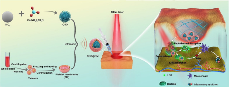
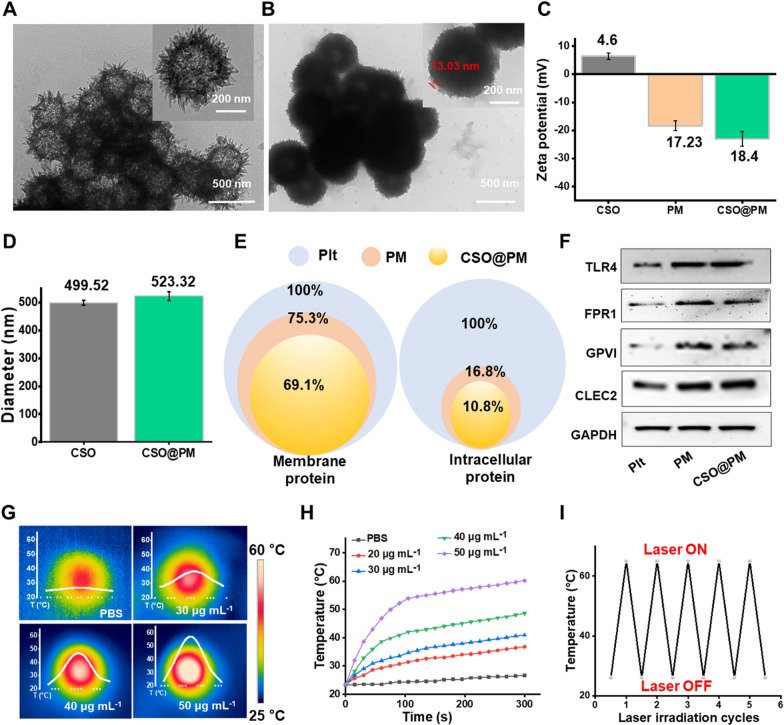
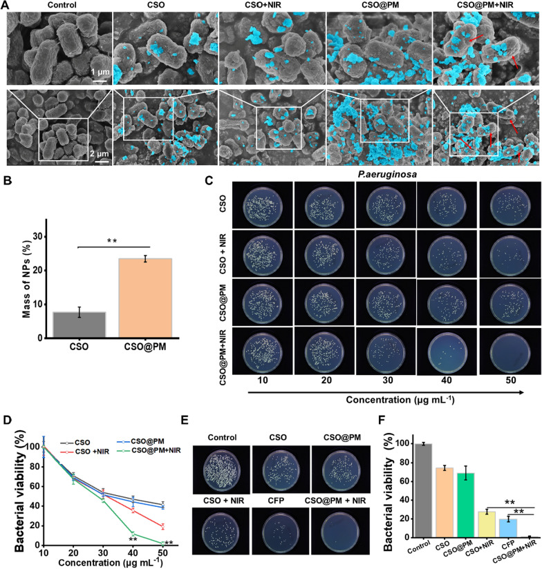
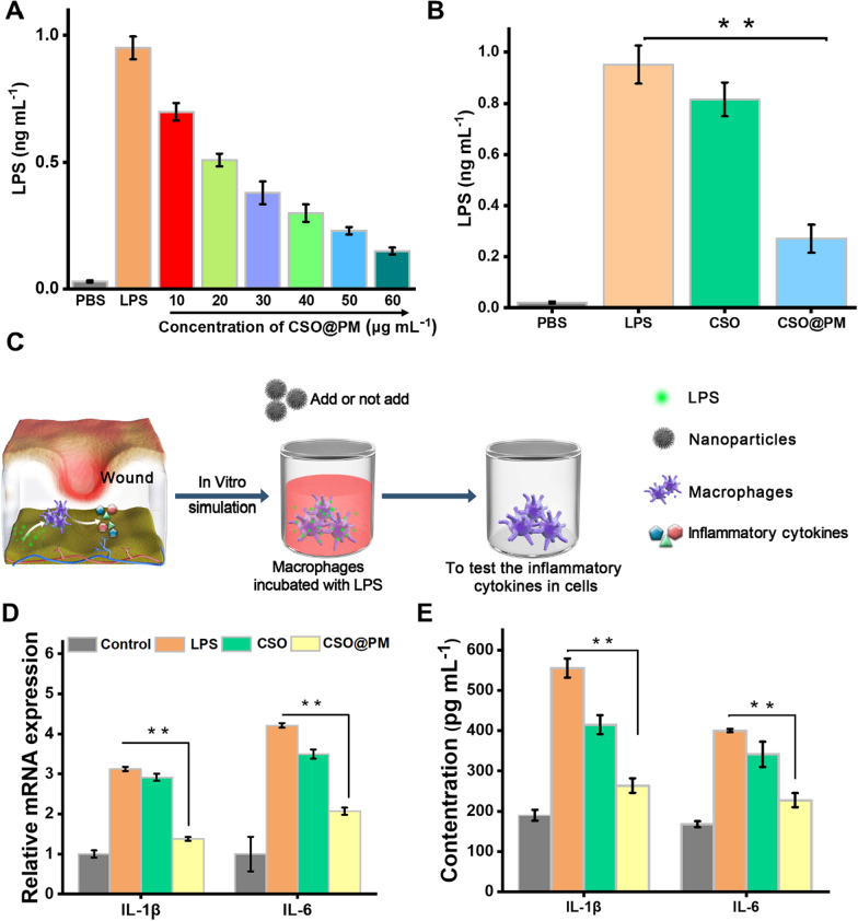
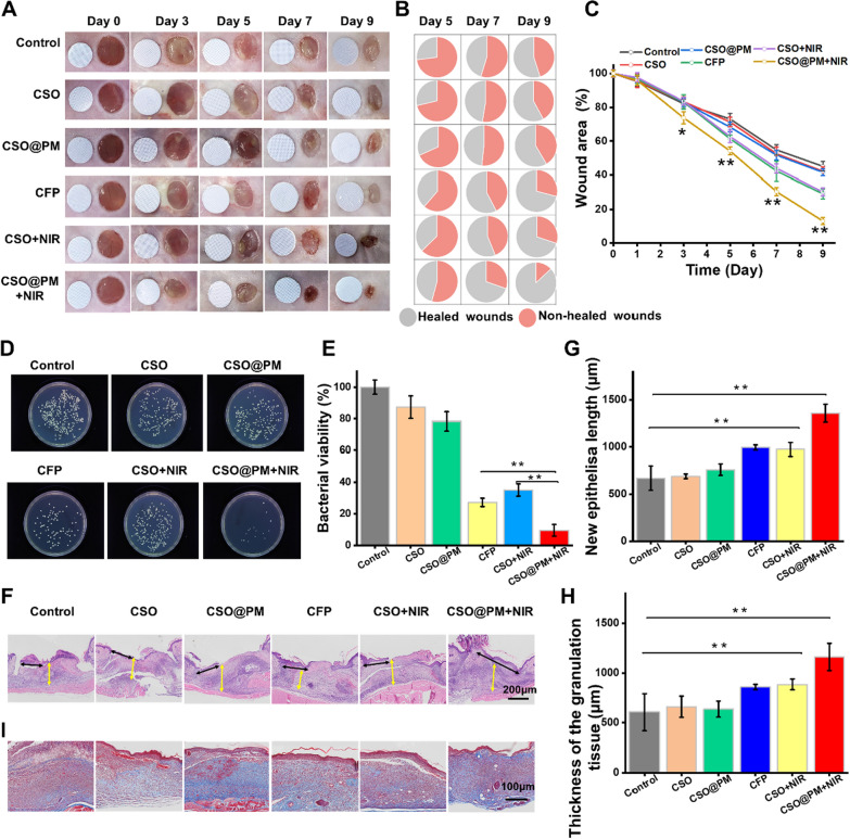
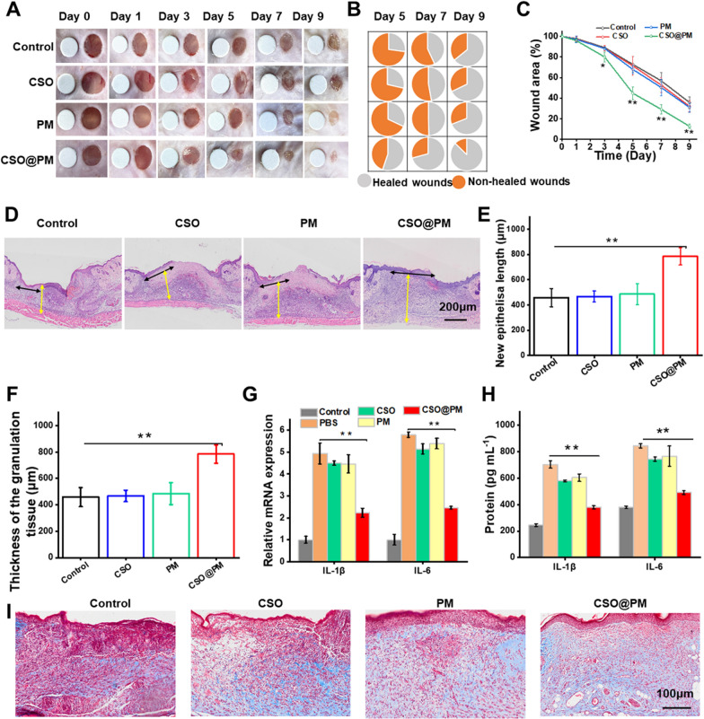
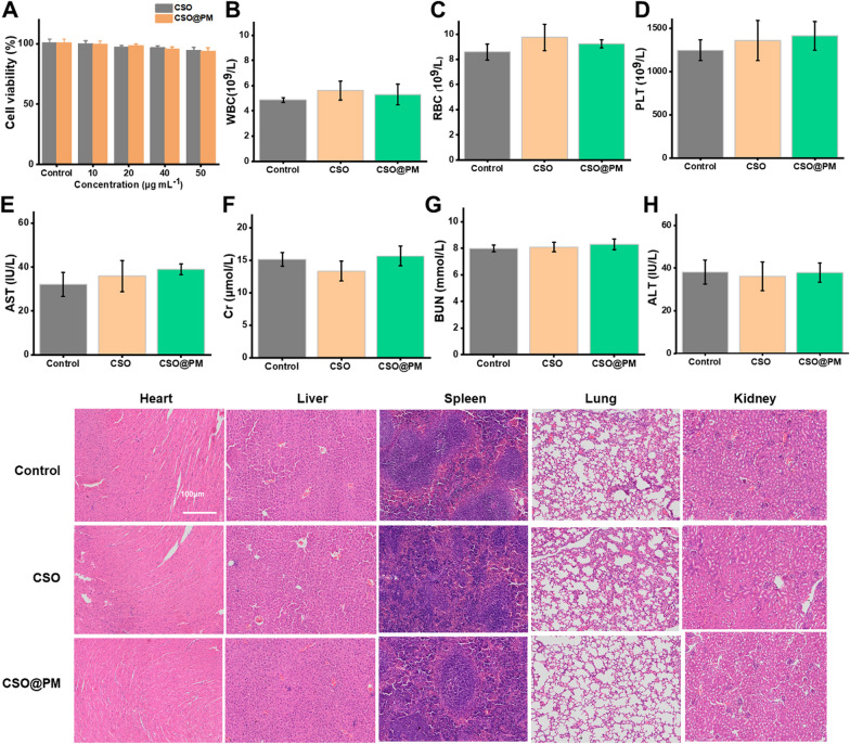
Serious infection caused by drug-resistant gram-negative bacteria and their secreted toxins (e.g., lipopolysaccharide) is a serious threat to human health. Thus, treatment strategies that efficiently kill bacteria and reducing the impact of their toxins simultaneously are urgently required. Herein, a novel antibacterial platform composed of a mesoporous copper silicate microsphere (CSO) core and a platelet membrane (PM) shell was prepared (CSO@PM). CSO@PM specifically targets bacteria owing to formyl peptide receptors on the PM and, combined with photothermal therapy (PTT), exhibits highly effective bacter icidal activity. Importantly, CSO@PM can adsorb lipopolysaccharide secreted by gram-negative bacteria, resulting in inflammation reduction. Thus, CSO@PM stimulates re-epithelialization and granulation-tissue formation, promoting wound healing. Moreover, this antibacterial platform exhibits no obvious toxicity at all the test concentrations in vitro and in vivo. Thus, CSO@PM exhibits a robust antibacterial effect and a strong toxin-adsorption capacity, facilitating the clinical treatment of many bacterial infections and the development of next-generation antibacterial nanoagents.

Keywords: Bacterial infection; Mesoporous copper silicate microspheres; Photothermal therapy; Toxin adsorption; Wound healing.


Conflict of interest statement

The authors declare that they have no competing interests.

Figures

Fig. 1
Fig. 1
Schematic showing the preparation of CSO@PM NPs and the treatment of wounds infected with multi-drug-resistant Gram-negative bacteria. CSO@PM NPs specifically target bacteria, adsorbing their LPS and, in combination with NIR laser irradiation, effectively kill them, reducing inflammatory reactions and ultimately promoting wound healing
Fig. 2
Fig. 2
Characterization of CSO@PM. Representative TEM images showing the morphologies of CSO A before and B after PM coating. C Zeta potential values for CSO, PM, and CSO@PM. Data are mean ± SD (n = 3). D Hydrodynamic diameters of CSO and CSO@PM. Data are mean ± SD (n = 3). E Preservation of the PM proteins and E intracellular proteins detected in platelets, PM, and CSO@PM using proteomic analysis. F Western blot analysis of TLR4, FRP1, GPVI, GLEC-2, and GAPDH in platelets, PM, and CSO@PM. G Corresponding thermographic images and H temperature evolution curves of CSO suspensions with different concentrations in PBS (pH 7.4) under 808 nm NIR laser irradiation at 1.5 W cm−2. I Thermal response of CSO under repeated laser irradiation (n = 5; 808 nm, 1.5 W cm–2)
Fig. 3
Fig. 3
In vitro bactericidal activity of CSO@PM. A SEM images for the control (PBS treated), CSO, CSO@PM, CSO + NIR, CSO@PM + NIR groups after incubation with P. aeruginosa. Blue indicates NPs. B Mass of NPs bound to P. aeruginosa after co-incubation with CSO or CSO@PM. Values are mean ± SD (n = 3) and ** indicates P < 0.001. C Representative images and D quantitative analysis of bacterial colonies formed by P. aeruginosa after exposure to CSO and CSO@PM with or without 808 nm NIR irradiation. The values are shown as mean ± SD (n = 3) and ** indicates P < 0.001 compared with the corresponding control group. E Representative images and F quantitative analysis of bacterial colonies formed by P. aeruginosa after exposure to CFP, CSO, or CSO@PM with or without 808 nm NIR irradiation. The values are shown as mean ± SD (n = 3) and ** indicates P < 0.001 compared with the control group
Fig. 4
Fig. 4
Anti-inflammatory activity of CSO@PM in vitro. A LPS adsorption capacity of CSO@PM. The initial concentration of LPS was 1 ng mL−1. B LPS adsorption for the PBS, LPS (100 ng mL−1), CSO (50 μg mL−1), and CSO@PM (50 μg mL−1) groups. C Schematic of the co-culture system composed of RAW246.7 cells, LPS, and CSO@PM. D mRNA and E protein expression of IL-1β and IL-6 in RAW264.7 macrophages. The values are shown as mean ± SD (n = 3) and ** indicates P < 0.001 compared with the corresponding control group
Fig. 5
Fig. 5
Effects of CSO@PM on the healing of P. aeruginosa-infected wounds. A Photographs of P. aeruginosa-infected wounds under different treatments. The round blue card with a 5 mm diameter indicates initial wound size. B Fractions of the wounds healed by the different treatments on days 5, 7, and 9. The values are shown as mean ± SD (n = 3). C Quantitative analysis of wound area for each group. The values are shown as mean ± SD (n = 3). * P < 0.1 and ** P < 0.001 compared to the control group. D Photographs and E quantitative counts of bacterial colonies formed by P. aeruginosa obtained from wound tissues. The values were shown as mean ± SD (n = 3). * P < 0.1 and ** P < 0.001 compared to the corresponding control group. F H&E staining images of mouse wound tissue from all groups at days 7. Black arrows indicate the length of newly regenerated epidermis. Yellow arrows indicate thickness of granulation tissue. Scale bar: 200 μm. G Neo-epidermis length and H thickness of granulation tissue data. The values are show as mean ± SD (n = 3). * P < 0.1 and ** P < 0.001 compared to the corresponding control group. I Masson images of mouse wound tissue from all groups on day 7. Scale bar: 100 μm
Fig. 6
Fig. 6
Effect of CSO@PM on LPS-infected wound healing. A Photographs of LPS-infected wounds under different treatments. The round blue card with a 5 mm diameter indicates initial wound size. B Fractions of the wounds healed by the different treatments on days 5, 7, and 9 (n = 3). C Quantitative wound area analysis for each group (n = 3). * P < 0.1 and ** P < 0.001 compared to the corresponding control group. D H&E images of mouse wound tissue from all groups at day 7. Black arrows indicate the length of newly regenerated epidermis. Yellow arrows indicate thickness of granulation tissue. Scale bar: 200 μm E Neo-epidermis length and F granulation tissue thickness data. G Real-time quantitative PCR detection results for IL-1β and IL-6 mRNA and H protein expression of IL-1β and IL-6 in LPS-infected wounds at day 7. The values are shown as mean ± SD (n = 3). * P < 0.1 and ** P < 0.001 compared to the corresponding control group. I Masson images of mouse wound tissue from all groups on day 7. Scale bar: 100 μm
Fig. 7
Fig. 7
In vitro and in vivo biocompatibility of CSO@PM. A Cell viabilities of 3T3 fibroblasts treated with different concentrations of CSO@PM. BH Hematology and blood biochemistry analysis results for healthy Balb/c mice sacrificed 21 days after intravenous injection with CSO@PM at a concentration of 500 mL−1 (n = 3). PBS-treated mice were used as controls. B White blood cells, C red blood cells, D platelets, E aspartate aminotransferase (AST), F creatinine (Cr), G blood urea nitrogen (BUN), and H alanine aminotransferase (ALT). I H&E staining images of the major organs (heart, liver, spleen, lung, and kidney) from mice 21 days after intravenous injection with CSO@PM (500 μg mL−1). Scale bar: 100 μm

Similar articles

Cited by

References

    1. Sugden R, Kelly R, Davies S. Combatting antimicrobial resistance globally. Nat Microbiol. 2016;1(10):16187. - PubMed
    1. Kaier K, Heister T, Götting T, Wolkewitz M, Mutters NT. Measuring the in-hospital costs of Pseudomonas aeruginosa pneumonia: methodology and results from a German teaching hospital. BMC Infect Dis. 2019;19(1):1028. - PMC - PubMed
    1. Herrera S, Bodro M, Soriano A. Predictors of multidrug resistant Pseudomonas aeruginosa involvement in bloodstream infections. Curr Opin Infect Dis. 2021. - PubMed
    1. Sirijatuphat R, Sripanidkulchai K, Boonyasiri A, Rattanaumpawan P, Supapueng O, Kiratisin P, et al. Implementation of global antimicrobial resistance surveillance system (GLASS) in patients with bacteremia. PLoS ONE. 2018;13(1):e0190132. - PMC - PubMed
    1. Zumla A, Azhar EI, Hui DS, Shafi S, Petersen E, et al. Global spread of antibiotic-resistant bacteria and mass-gathering religious events. Lancet Infect Dis. 2018;18(5):488–490. - PubMed

MeSH terms