. 2021 Nov 22;19(1):385.
doi: 10.1186/s12951-021-01133-7.

Hydroxypropylmethylcellulose as a film and hydrogel carrier for ACP nanoprecursors to deliver biomimetic mineralization

Affiliations

Hydroxypropylmethylcellulose as a film and hydrogel carrier for ACP nanoprecursors to deliver biomimetic mineralization

Zhe Wang et al. J Nanobiotechnology. .

Erratum in

Abstract

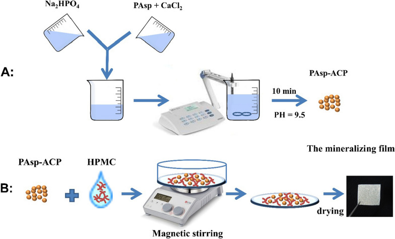
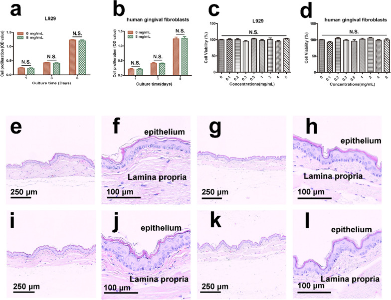
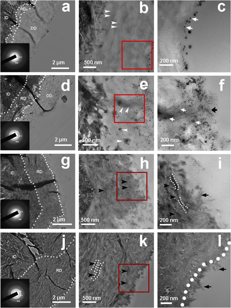
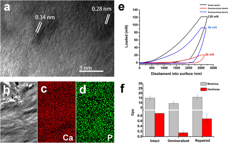
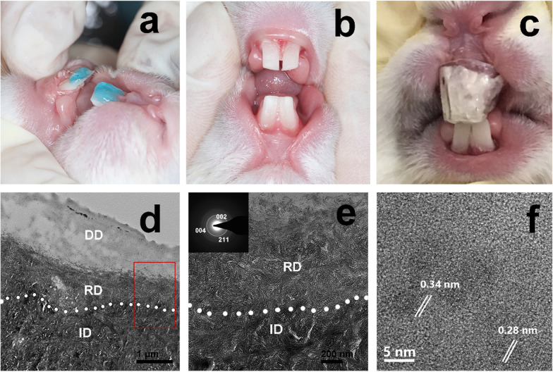
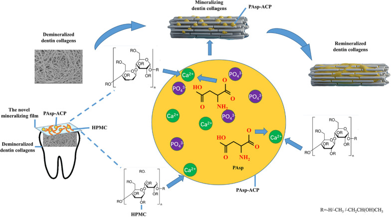
Demineralization of hard tooth tissues leads to dental caries, which cause health problems and economic burdens throughout the world. A biomimetic mineralization strategy is expected to reverse early dental caries. Commercially available anti-carious mineralizing products lead to inconclusive clinical results because they cannot continuously replenish the required calcium and phosphate resources. Herein, we prepared a mineralizing film consisting of hydroxypropylmethylcellulose (HPMC) and polyaspartic acid-stabilized amorphous calcium phosphate (PAsp-ACP) nanoparticles. HPMC which contains multiple hydroxyl groups is a film-forming material that can be desiccated to form a dry film. In a moist environment, this film gradually changes into a gel. HPMC was used as the carrier of PAsp-ACP nanoparticles to deliver biomimetic mineralization. Our results indicated that the hydroxyl and methoxyl groups of HPMC could assist the stability of PAsp-ACP nanoparticles and maintain their biomimetic mineralization activity. The results further demonstrated that the bioinspired mineralizing film induced the early mineralization of demineralized dentin after 24 h with increasing mineralization of the whole demineralized dentin (3-4 µm) after 72-96 h. Furthermore, these results were achieved without any cytotoxicity or mucosa irritation. Therefore, this mineralizing film shows promise for use in preventive dentistry due to its efficient mineralization capability.

Keywords: Biomimetic mineralization; Collagen; Dental caries; Dentin; Film; Hydroxypropylmethylcellulose.


Conflict of interest statement

The authors declare that they have no competing interests.

Figures

Fig. 1
Fig. 1
Schematic diagram showing the preparation of the mineralizing film. A Preparation of the PAsp-ACP nanoparticles. B Preparation of the mineralizing film
Fig. 2
Fig. 2
Characterization of ACP (a1a4), PAsp-ACP nanoparticles (b1b4) and the mineralizing film (c1c4). a1, b1 SEM images of ACP and PAsp nanoparticles show the particles were spherical. a2, b2 TEM images of ACP and PAsp nanoparticles show the diameter of an individual nanoparticle is approximately 30–80 nm. a3, b3 FTIR spectra of ACP and PAsp nanoparticles shows characteristic amorphous peaks at 1050 cm−1 and 580 cm−1. a4, b4 XRD patterns of the ACP and PAsp-ACP nanoparticles show the broad peak, the peak in a4 is a small left offset compared to that of b4 and c4. c1 SEM image shows that the PAsp-ACP nanoparticles are homogeneously distributed in the mineralizing film. c2 TEM image shows that the spherical PAsp-ACP nanoparticles are dispersed in HPMC, and the SAED pattern (inset panel) indicates that the PAsp-ACP nanoparticles are amorphous. c3 FTIR spectra shows that HPMC exhibits a typical C–O peak at 1065 cm−1, C–O–C peak at 1119 cm−1, and CO32− peaks at 872 cm−1 and 1420 cm−1 (black line). The PAsp-ACP nanoparticles exhibit characteristic PO43− absorption peaks at 1050 cm−1 and 580 cm−1 as well as absorption peaks attributed to bound water at 1300 cm−1 and 1750 cm−1 (red line). The mineralizing film exhibits characteristic peaks of both HPMC and the ACP nanoparticles (blue line). c4 The XRD pattern of the mineralizing film displays a broad peak at 2θ = 30°
Fig. 3
Fig. 3
Ca and P release and phase transformations. a Release kinetics for calcium (black line) and phosphate (red line) ions from the mineralizing film over 24 h. b FTIR spectra of the mineralizing film in AS show a peak at 580 cm−1 within 6 h and two peaks at 560 cm−1 and 600 cm−1 over 8–48 h. c Magnified images of the spectral curve ranging from 400 to 750 cm−1 of b. d SF scheme. A1/A2 = 0 (noncrystallization) to 1 (complete crystallization). e Kinetics of the ACP to HAp phase transformation
Fig. 4
Fig. 4
Cryo-TEM images of the mineralizing film and its SAED pattern at 0 h (a, f), 6 h (b, g), 8 h (c, h), 12 h (d, i) and 24 h (e, j) in artificial saliva. a, b The spherical ACP nanoparticles in the mineralizing film are approximately 30–80 nm in diameter and stable in artificial saliva over 6 h. c The ACP nanoparticles decreased in size and fused at 8 h. d, e Most of the ACP nanoparticles transformed into HAp at 12 and 24 h. fj SAED patterns of the mineralizing film
Fig. 5
Fig. 5
Cytotoxicity and oral mucosa irritation tests of the mineralizing film. ad CCK-8 assay of L929 and human gingival fibroblasts. el Histological sections of oral mucosa from golden hamster cheek pouches. The oral mucosa was treated either with polar (0.9% NaCl) and nonpolar (cottonseed oil) liquid in the control group (eh), or with the polar and nonpolar extracts of mineralizing film in the experimental group (il). f, h, j, and l are magnified images of e, g, i, and k. The stratified squamous epithelium and lamina propria were in normal arrangement. No cell proliferation, no edema, no inflammatory cells and no cell necrosis were detected
Fig. 6
Fig. 6
TEM images of dentin treated with the mineralizing film for 24 h (ac), 48 h (df), 72 h (gi) and 96 h (jl). c, f, i and l are magnified images of b, e, h and k, respectively. At 24 h, spherical ACP nanoparticles were attached to the surface (c, white arrow). At 48 h, some nanoparticles (f, black arrow) were observed on the surface, and some were observed in the middle of the demineralized dentin layer (e, f, white arrow). The collagens became thicker and darker (e, white arrow). Rod-like crystals were detected on the surface of the remineralized dentin after 72 h (i, black arrow), and the demineralized dentin was fully mineralized and fused with the surface crystals of the dentin. A needle-like HAp layer was detected on the dentin surface at 96 h (l, black arrow). Both the black arrow and white dotted line (k, l) indicate the remineralized dentin collagen. ID, intact dentin; DD, demineralized dentin; RD, remineralized dentin
Fig. 7
Fig. 7
HRTEM images, elemental maps and nanoindentation tests of the dentin. a HRTEM image shows the two interplanar spacings: 0.34 nm and 0.28 nm. b Remineralized dentin. c, d Elemental maps revealing the uniform distribution of calcium and phosphate of b. e Load–displacement curves show the different load forces exerted on the intact dentin, demineralized dentin and remineralized dentin at the same dent depth (2500 nm). f Hardness and elastic modulus values for the intact dentin, demineralized dentin and remineralized dentin. Both the remineralized and intact dentin exhibited much higher values than the demineralized dentin
Fig. 8
Fig. 8
In vivo remineralization experiment of demineralized dentin of rabbits. a The rabbit dentins were etching by 37% phosphoric acid for 15 s. b The demineralized dentins were obtained after rinsing and gentle drying. c The mineralizing film attached on the demineralized dentin surface was covered with a transparent customized tray. df TEM images of the demineralized dentin treated with the mineralizing film for 7 days show that the remineralized layer was approximately 600 nm thick [d, the magnified images of d (e, f)]. The SAED patterns (insets in e) reveal 002, 004, and 211 diffraction rings. f The HRTEM image shows two interplanar spacings: 0.34 nm and 0.28 nm. ID, intact dentin; DD, demineralized dentin; RD, remineralized dentin
Fig. 9
Fig. 9
Diagram showing the mechanism of the remineralization of dentin collagens. Once in contact with water, the mineralizing film attaches to the demineralized dentin. The large number of hydroxyl, methyl and methoxy anion groups of the HPMC gel crosslink with calcium ions in synergy with PAsp. Finally, they stabilize the ACP precursors and induce the remineralization of dentin collagens

Similar articles

Cited by

References

    1. Peres MA, Macpherson LMD, Weyant RJ, Daly B, Venturelli R, Mathur MR, et al. Oral diseases: a global public health challenge. Lancet. 2019;394(10194):249–260. - PubMed
    1. Bernabé E, Marcenes W. Can minimal intervention dentistry help in tackling the global burden of untreated dental caries? Br Dent J. 2020;229(7):487–491. - PubMed
    1. Merchan MT, Ismail AI. Measurement and distribution of dental caries. In: Burt and Eklund’s dentistry, dental practice, and the community. Seventh Edition. 2021. p. 154–70. 10.1016/b978-0-323-55484-8.00014-9.
    1. Vanaei S, Parizi MS, Vanaei S, Salemizadehparizi F, Vanaei HR. An overview on materials and techniques in 3D bioprinting toward biomedical application. Eng Regener. 2021;2:1–18.
    1. Feng X, Hou X, Cui C, Sun S, Sadik S, Wu S, et al. Mechanical and antibacterial properties of tannic acid-encapsulated carboxymethyl chitosan/polyvinyl alcohol hydrogels. Eng Regener. 2021;2:57–62.

MeSH terms