. 2021 Nov 22;21(1):379.
doi: 10.1186/s12890-021-01751-9.

The function role of ubiquitin proteasome pathway in the ER stress-induced AECII apoptosis during hyperoxia exposure

Affiliations

The function role of ubiquitin proteasome pathway in the ER stress-induced AECII apoptosis during hyperoxia exposure

Yue Zhu et al. BMC Pulm Med. .

Abstract

Background: Bronchopulmonary dysplasia (BPD) is a major cause of mortality and morbidity in premature infants, characterized by alveolar dysplasia and pulmonary microvascular remodeling. In the present study, we have investigated the functional roles of ubiquitin proteasome pathway (UPP) in BPD, and its relationship with endoplasmic reticulum stress (ERS) mediated type II alveolar epithelial cell (AECII) apoptosis.

Methods: A hyperoxia-induced BPD rat model was constructed and the pathologic changes of lung tissues were evaluated by hematoxylin-eosin staining. Cell apoptosis and protein expression were determined by TUNEL assay and Western blotting, respectively. Further reagent kit with specific fluorescent substrate was utilized to measure the activity of 20 s proteasome. Meanwhile, AECII were cultured in vitro and exposed to hyperoxia. AECII apoptosis were measured by flow cytometry. In contrast, MG132 treatment was induced to explore UPP during hyperoxia exposure on AECII apoptosis and ERS sensors expression.

Results: A significant increase in apoptosis and total ubiquitinated proteins expression were observed in BPD rats and AECII culture, and the change of UPP was associated with ERS. In order to confirm the role of UPP in AECII apoptosis of BPD, AECII cells were treated by MG132 with the concentration of 10 μmol/L under hyperoxia exposure. We found that the proteins expression of glucose-regulated protein 78 (GRP-78), PKR-like ER kinase (PERK), activating transcription factor 4 (ATF4), activating transcription factor 6 (ATF6) and C/EBP homologous protein (CHOP), as well as AECII apoptosis were increased following MG132 treatment. Furthermore, the relatively up-regulated in the levels of total ubiquitinated proteins expression and 20 s proteasome activity were correlated with increased ERS sensors expression.

Conclusions: Our findings indicate that UPP may participate in the ERS-induced AECII apoptosis under hyperoxia condition.

Keywords: Alveolar epithelial type II cells; Apoptosis; Bronchopulmonary dysplasia; Endoplasmic reticulum stress; Ubiquitin proteasome pathway.


Conflict of interest statement

Not applicable.

Figures
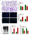
Fig. 1
Fig. 1
Apoptosis and Total ubiquitinated proteins expression in BPD rats. Neonatal rats were killed at P 7 and P14; normoxia group: 21% O2, hyperoxia group: 8085% O2. A HE staining assay were used to observe the morphological changes of lung tissue and showed a significant decrease in radial alveolar count (Scale bar = 10 µm; Original magnifications: × 100). B TUNEL assay demonstrated a significant increase in the apoptosis index, in a pattern similar to that observed by immunofluorescence staining (Scale bar = 20 µm; Original magnifications: × 200; square frame magnification: × 400). C The expression of ubiquitinated proteins were detected by Western blot assays in the four groups and the grouping of blots cropped from the same gel. β-actin was used as the loading control. The part of original data for were presented in Additional file 1: Fig. S1. D Reagent kit with specific fluorescent substrate was utilized to measure the activity of 20 s proteasome. Values represent mean ± SD; ** P < 0.05 versus normoxia group
Fig. 2
Fig. 2
Hyperoxia caused AECII apoptosis and ubiquitin proteasome pathway changed. A After 24, 48 and 72 h hyperoxia exposure, AECII apoptosis was analyzed by Annexin V-FITC/PI double staining followed by flow cytometry. B The expression of ubiquitinated proteins were detected by Western blot assays in different groups and the grouping of gels cropped from the same gel. β-actin was used as the loading control. The part of original data were presented in Additional file 1: Fig. S2. C Reagent kit with specific fluorescent substrate was utilized to measure the activity of 20 s proteasome. Values represent mean ± SD; ** P < 0.05 versus normoxia group
Fig. 3
Fig. 3
Effects of the protein expression of GRP-78, PERK, ATF4, ATF6 and CHOP in AECII after hyperoxia exposure. A Western blot analysis of the AECII that were exposed to 95% oxygen (hyperoxia) or not (normoxia) for 24, 48 and 72 h, 30 g of total protein per well were loaded and subjected to Western blot analysis with corresponding antibodies shown on the left. The part of original data were presented in Additional file 1: Fig. S3. BF Quantification of signals on the Fig. 3A, the results showed an increase in GRP78, PERK, ATF4, ATF6 and CHOP protein expression in hyperoxia group in comparison with normoxia group in 24, 48, and 72 h. Values are presented as mean ± SD; ** P < 0.05 versus normoxia group
Fig. 4
Fig. 4
Effects of MG132 treatment on ubiquitin proteasome pathway in AECII during hyperoxia exposure. After MG132 treatment with varying concentration (0, 5, 10, 15 and 50 μmol/L), cell viability was detected by MTT assay (A) and the activity of 20 s proteasome was detected by Reagent kit with specific fluorescent substrate (B). Compared with Control group and DMSO group, MG132 group (MG132 concentration of 10 μmol/L) showed a decreased activity of 20 s proteasome (C) and an increased expression of total ubiquitinated proteins (D). The grouping of blots cropped from different parts of the same gel and β-actin was used as the loading control. The part of original data of D were presented in Additional file 1: Fig. S4. Values represent mean ± SD; ** P < 0.05 versus control
Fig. 5
Fig. 5
Effects of MG132 treatment on AECII apoptosis during hyperoxia exposure. a After MG132 treatment, AECII apoptosis was analyzed by Annexin V-FITC/PI double staining followed by flow cytometry. b The proteins expression of Caspases-3, Bax and Bcl-2 were detected by Western blot assays, as well as quantification of Caspases-3 and ratio of Bax/Bcl-2 was measured in different groups. β-actin was used as the loading control. The part of original data were presented in Additional file 1: Fig. S5. Values represent mean ± SD; ** P < 0.05 versus control
Fig. 6
Fig. 6
Effects of MG132 treatment on GRP-78, PERK, ATF4, ATF6 and CHOP expression during hyperoxia exposure. a Western blot analysis of the AECII that were exposed to hyperoxia for 72 h, 30 g of total protein per well were loaded and subjected to Western blot analysis with corresponding antibodies shown on the left. The part of original data were presented in Additional file 1: Fig. S6. BF Quantification of signals on the A, the results showed an increase in GRP78, PERK, ATF4, ATF6 and CHOP protein expression in hyperoxia group in comparison with normoxia group in 72 h. Values are presented as mean ± SD; ** P < 0.05 versus control

Similar articles

Cited by

References

    1. Thébaud B, Goss KN, Laughon M, Whitsett JA, Abman SH, Steinhorn RH, Aschner JL, Davis PG, McGrath-Morrow SA, Soll RF, et al. Bronchopulmonary dysplasia. Nat Rev Dis Primers. 2019;5(1):78. - PMC - PubMed
    1. Tanaka T, Saito Y, Matsuda K, Kamio K, Abe S, Kubota K, Azuma A, Gemma A. Cyclic mechanical stretch-induced oxidative stress occurs via a NOX-dependent mechanism in type II alveolar epithelial cells. Respir Physiol Neurobiol. 2017;242:108–116. - PubMed
    1. McAdams RM, Mustafa SB, Shenberger JS, Dixon PS, Henson BM, DiGeronimo RJ. Cyclic stretch attenuates effects of hyperoxia on cell proliferation and viability in human alveolar epithelial cells. Am J Physiol Lung Cell Mol Physiol. 2006;291(2):L166–L174. - PMC - PubMed
    1. Jin L, Yang H, Fu J, Xue X, Yao L, Qiao L. Association between oxidative DNA damage and the expression of 8-oxoguanine DNA glycosylase 1 in lung epithelial cells of neonatal rats exposed to hyperoxia. Mol Med Rep. 2015;11(6):4079–4086. - PMC - PubMed
    1. Lozon TI, Eastman AJ, Matute-Bello G, Chen P, Hallstrand TS, Altemeier WA. PKR-dependent CHOP induction limits hyperoxia-induced lung injury. Am J Physiol Lung Cell Mol Physiol. 2011;300(3):L422–429. - PMC - PubMed

MeSH terms