. 2021 Nov 30;118(48):e2111247118.
doi: 10.1073/pnas.2111247118.

Claudin-2 and claudin-12 form independent, complementary pores required to maintain calcium homeostasis

Affiliations

Claudin-2 and claudin-12 form independent, complementary pores required to maintain calcium homeostasis

Megan R Beggs et al. Proc Natl Acad Sci U S A. .

Abstract

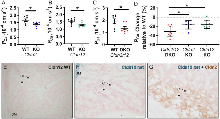
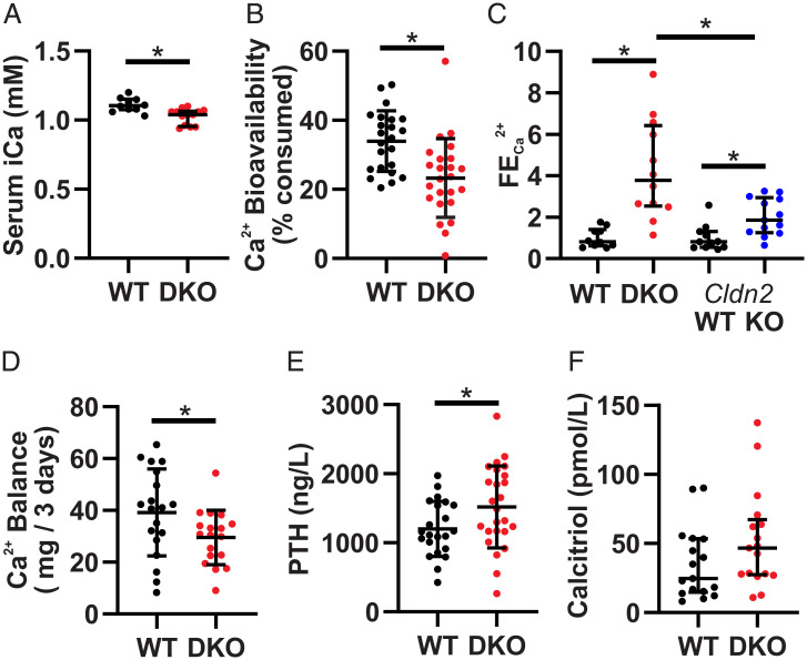
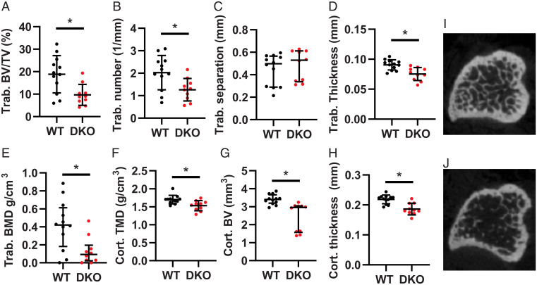
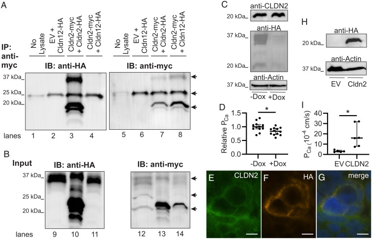
Calcium (Ca2+) homeostasis is maintained through coordination between intestinal absorption, renal reabsorption, and bone remodeling. Intestinal and renal (re)absorption occurs via transcellular and paracellular pathways. The latter contributes the bulk of (re)absorption under conditions of adequate intake. Epithelial paracellular permeability is conferred by tight-junction proteins called claudins. However, the molecular identity of the paracellular Ca2+ pore remains to be delineated. Claudins (Cldn)-2 and -12 confer Ca2+ permeability, but deletion of either claudin does not result in a negative Ca2+ balance or increased calciotropic hormone levels, suggesting the existence of additional transport pathways or parallel roles for the two claudins. To test this, we generated a Cldn2/12 double knockout mouse (DKO). These animals have reduced intestinal Ca2+ absorption. Colonic Ca2+ permeability is also reduced in DKO mice and significantly lower than single-null animals, while small intestine Ca2+ permeability is unaltered. The DKO mice display significantly greater urinary Ca2+ wasting than Cldn2 null animals. These perturbations lead to hypocalcemia and reduced bone mineral density, which was not observed in single-KO animals. Both claudins were localized to colonic epithelial crypts and renal proximal tubule cells, but they do not physically interact in vitro. Overexpression of either claudin increased Ca2+ permeability in cell models with endogenous expression of the other claudin. We find claudin-2 and claudin-12 form partially redundant, independent Ca2+ permeable pores in renal and colonic epithelia that enable paracellular Ca2+ (re)absorption in these segments, with either one sufficient to maintain Ca2+ balance.

Keywords: calcium; claudins; paracellular.


Conflict of interest statement

The authors declare no competing interest.

Figures

Fig. 1.
Fig. 1.
Cldn2 and Cldn12 confer independent Ca2+ permeability to the proximal colon. PCa measured ex vivo in Ussing’s chambers across the proximal colon compared to WT of Cldn2 KO (A) (n = 8 per group and P = 0.016); Cldn12 KO (B) (n = 8 per group and P = 0.012); and Cldn2/12 DKO (C) (n = 8 per group and P = 0.019). Full results of bionic dilution potential experiments on small intestine and colon of DKO mice are in SI Appendix, Fig. S2 and Tables S1–S4. Means were compared by Student’s t test. (D) Data from A–C expressed as the percentage change in PCa relative to WT for each genotype. Data are presented as mean ± SD. One-way ANOVA with Dunnett correction for multiple comparisons to compare mean from DKO mice to Cldn2 KO (P = 0.017) and Cldn12 KO (P = 0.010) was performed. The Cldn12 coding exon was replaced with a LacZ cassette. (E and F) We used this to localize Cldn12 expression by X-gal staining of colon from Cldn12 WT (E) and Cldn12 heterozygous mice (F). Staining is present in colonic crypt epithelial cells in Cldn12 heterozygous mouse (cyan). (Scale bars, 100 µm [E] and 25 µm [F]). (G) X-gal staining of colon for Cldn12 (cyan) and immunohistochemical staining for claudin-2 (brown) from a Cldn12 heterozygous mouse. Cr = crypts, SM = smooth muscle, and L = colonic lumen. (Scale bar, 25 µm.) *P < 0.05.
Fig. 2.
Fig. 2.
Cldn2/12 DKO mice have hypocalcemia, hypercalciuria, and decreased intestinal Ca2+ absorption. (A) Serum-ionized Ca2+ (median ± interquartile range [IQR], n = 10 WT, 13 DKO, Mann–Whitney U Test, and P = 0.002). (B) Ca2+ bioavailability as a percent of Ca2+ consumed (mean ± SD, n = 23 WT, 24 DKO, Student’s t test, and P = 0.001). (C) Fractional excretion of urinary Ca2+ normalized to WT for each genotype (median ± IQR, n = 10 WT, 12 DKO, 11 Cldn2 WT, 13 Cldn2 KO, Mann–Whitney U test, P < 0.0001 WT versus DKO, P = 0.003 Cldn2 WT versus KO, and P = 0.011 DKO versus Cldn2 KO). (D) Net 3 d Ca2+ balance (mean ± SD, n = 19 WT, 18 DKO, Student’s t test, and P = 0.043). (E) Serum PTH levels (mean ± SD, n = 22 WT, 25 DKO, Student’s t test, and P = 0.039). (F) Serum calcitriol (median ± IQR, n = 17 WT, 17 DKO, Mann–Whitney U Test, and P = 0.099). *P < 0.05.
Fig. 3.
Fig. 3.
Cldn2/12 DKO mice have altered bone morphometry at 3 mo. Microarchitecture of trabecular (trab) (A–E) and cortical (cort) (FH) bone from tibia of WT and Cldn2/12 DKO mice analyzed by micro-CT. (A) Trabecular bone volume/tissue volume (P = 0.006). (B) Trabecular number (P = 0.014). (C) Trabecular separation (P = 0.228). (D) Trabecular thickness (P = 0.001). (E) Trabecular bone mineral density (P = 0.014). (F) Cortical tissue mineral density (P = 0.006). (G) Cortical bone volume (P = 0.0001). (H) Cortical thickness (P = 0.0003). Representative micro-CT images of the tibial metaphyses shown at 40 slices from growth plate from WT (I) and DKO (J) mice. Data are presented as mean ± SD compared by unpaired t test (A, B, D, G, and H) or as median (IQR) compared by Mann–Whitney U test (C, E, and F). *P < 0.05.
Fig. 4.
Fig. 4.
Claudin-2 and claudin-12 form separate Ca2+ permeable pores. (A) HEK293 cells transfected as indicated were lysed or the buffer without cell lysate as control (no lysate lane) and proteins immunoprecipitated (IP) with antibodies to myc before sodium dodecyl sulphate–polyacrylamide gel electrophoresis (SDS-PAGE). Proteins were electrotransferred and blotted with anti-HA antibody (lanes 1 through 4) and then stripped and reprobed with an anti-myc antibody (lanes 5 through 8). IB, immunoblot. (B) Protein from HEK293 cells above directly resolved on SDS-PAGE (Input) and blotted with anti-HA (lanes 9 through 11) and then anti-myc (lanes 12 through 14). Claudin dimers (37 kDa) and monomers (20 kDa) are highlighted by arrows. The arrow at 25 kDa illustrates IgG light chain in the IP samples as seen in the lane with no cell lysate added. EV, empty vector. Representative blot of three repeats is shown. (C) Caco-2 cells endogenously express CLDN2 and express HA-tagged CLDN12 via a tet-off system. (D) Caco-2 cells expressing CLDN2 and CLDN12 (−Dox) have greater Ca2+ permeability than cells expressing only CLDN2 (+Dox) (P = 0.014 and unpaired t test). (EG) Immunofluorescence of Caco-2 cells for endogenous CLDN2 (green) and HA-tagged CLDN12 (orange) demonstrating colocalization of CLDN2 and CLDN12 in the same cell membrane. (H) Opossum kidney (OK) cells endogenously expressing CLDN12 and expressing either EV or CLDN2. (I) OK cells expressing CLDN2 and CLDN12 (“CLDN2” data) have greater Ca2+ permeability than cells expressing only CLDN12 (“EV” data) (P = 0.018, Welch’s t test). Dox, doxycycline. TER, pNa, pCl, and relative permeabilities are presented in SI Appendix, Tables S8 and S9. *P < 0.05.

References

    1. Hoenderop J. G. J., Nilius B., Bindels R. J. M., Calcium absorption across epithelia. Physiol. Rev. 85, 373–422 (2005). - PubMed
    1. Klibanski A., et al. , NIH Consensus Development Panel on Osteoporosis Prevention, Diagnosis, and Therapy, Osteoporosis prevention, diagnosis, and therapy. JAMA 285, 785–795 (2001). - PubMed
    1. Johnell O., Kanis J. A., An estimate of the worldwide prevalence and disability associated with osteoporotic fractures. Osteoporos. Int. 17, 1726–1733 (2006). - PubMed
    1. Plain A., Alexander R. T., Claudins and nephrolithiasis. Curr. Opin. Nephrol. Hypertens. 27, 268–276 (2018). - PubMed
    1. Christakos S., et al. , Vitamin D and the intestine: Review and update. J. Steroid Biochem. Mol. Biol. 196, 105501 (2020). - PMC - PubMed

Publication types

LinkOut - more resources