. 2021 Nov 22;11(1):22654.
doi: 10.1038/s41598-021-01809-y.

The evolutionary conserved TLDc domain defines a new class of (H+)V-ATPase interacting proteins

Affiliations

The evolutionary conserved TLDc domain defines a new class of (H+)V-ATPase interacting proteins

A F Eaton et al. Sci Rep. .

Erratum in

Abstract

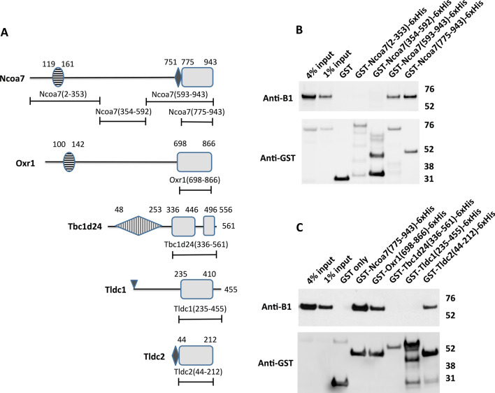
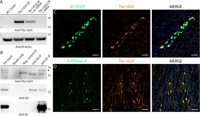
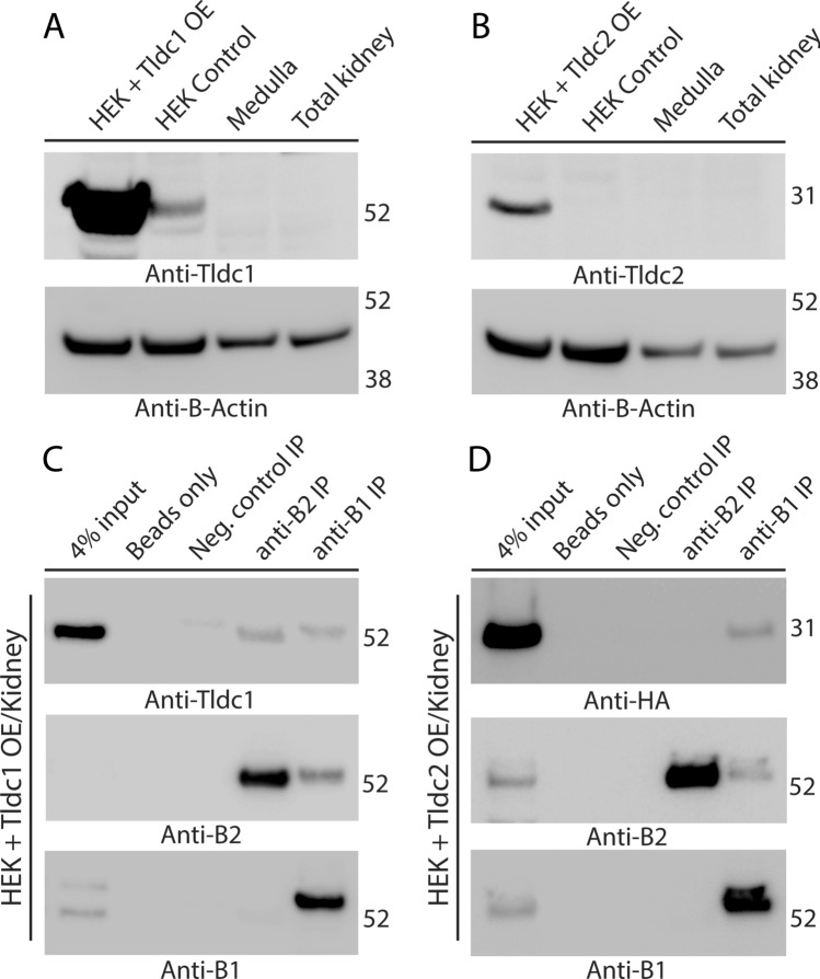
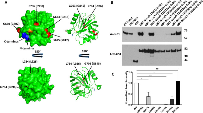
We recently found that nuclear receptor coactivator 7 (Ncoa7) and Oxr1 interact with the proton-pumping V-ATPase. Ncoa7 and Oxr1 belong to a group of proteins playing a role in the oxidative stress response, that contain the conserved "TLDc" domain. Here we asked if the three other proteins in this family, i.e., Tbc1d24, Tldc1 and Tldc2 also interact with the V-ATPase and if the TLDc domains are involved in all these interactions. By co-immunoprecipitation, endogenous kidney Tbc1d24 (and Ncoa7 and Oxr1) and overexpressed Tldc1 and Tldc2, all interacted with the V-ATPase. In addition, purified TLDc domains of Ncoa7, Oxr1 and Tldc2 (but not Tbc1d24 or Tldc1) interacted with V-ATPase in GST pull-downs. At the amino acid level, point mutations G815A, G845A and G896A in conserved regions of the Ncoa7 TLDc domain abolished interaction with the V-ATPase, and S817A, L926A and E938A mutations resulted in decreased interaction. Furthermore, poly-E motifs upstream of the TLDc domain in Ncoa7 and Tldc2 show a (nonsignificant) trend towards enhancing the interaction with V-ATPase. Our principal finding is that all five members of the TLDc family of proteins interact with the V-ATPase. We conclude that the TLDc motif defines a new class of V-ATPase interacting regulatory proteins.


Conflict of interest statement

The authors declare no competing interests.

Figures

Figure 1
Figure 1
The TLDc domain is sufficient to mediate interaction between TLDc protein family members Ncoa7, Oxr1 and Tldc2 and the kidney-specific B1 subunit of V-ATPase in GST pull-down assay. (A) Schematic representation of domain architecture of the TLDc proteins and constructs used to study the role of the conserved TLDc domain in the interaction with V-ATPase. Boundaries of the domains and constructs are indicated as amino acid numbers, as in the longest known isoform of the corresponding mouse protein. The conserved regions of TLDc domains are shown as grey rectangles, the non-conserved loop in the TLDc domain of Tbc1d24 and C-terminal extensions in the TLDc domains of both Tbc1d24 and Tldc1 are shown as lines. The horizontally striped ovals are LysM domains in Ncoa7 and Oxr1. The small black rhombuses in Ncoa7 and Tldc2 are poly-E rich motifs. The vertically striped rhombus in Tbc1d24 is a TBC domain. The small triangle at the beginning of Tldc1 indicates its site of myristylation. (B) Anti-B1 and anti-GST western blots of a representative GST pull-down assay using the purified GST-tagged N-terminal region of Ncoa7 (2–353), the middle region of Ncoa7 (354–592), the C-terminal region of Ncoa7 (593–943), and the Ncoa7 TLDc domain (775–943) as baits and kidney lysate containing the endogenous B1 subunit of V-ATPase as a prey. Numbers in parentheses indicate the amino acid boundaries of the constructs, based on of the longest known mouse isoform of Ncoa7. This experiment was repeated five times with similar results. (C) Anti-B1 and anti-GST western blots of a representative GST pull-down assay, using the purified GST-tagged TLDc domains of Ncoa7 (775–943), Oxr1 (698–866), Tbc1d24 (336–561), Tldc1 (235–455) and Tldc2 (44–212) and kidney lysate as the source of the B1 subunit of V-ATPase. In both panels (B) and (C) GST only pull-down was used as a negative control; anti-GST blot was used as a loading control for comparison between samples. This experiment was repeated three times with similar results.
Figure 2
Figure 2
Deletion of a non-conserved loop in the TLDc domain of Tbc1d24 and the C-terminal extensions of both Tbc1d24 and Tldc1, does not result in their interaction with V-ATPase in GST pull down assay. The GST-tagged full-length purified recombinant Tldc1 (2–455) does not interact with V-ATPase in GST pull-down assay. (A) Schematic representation of the domain architecture of Tbc1d24 and Tldc1 proteins and the constructs used to study the role of the non-conserved insertion in Tbc1d24 and the C-terminal extensions in both Tbc1d24 and Tldc1 in their interaction with V-ATPase. Boundaries of the domains, non-conserved regions and constructs are indicated as in Fig. 1. (B) Anti-B1 and anti-GST western blots of a representative GST pull-down assay, using the purified GST-tagged TLDc domains of Tbc1d24 (336–561) and Tldc1 (235–455), as well as, the GST-tagged truncated versions of the TLDc domains of Tbc1d24 and Tldc1: Tbc1d24 (336-446_496-556) and Tldc1 (235–410), and the GST-tagged full-length recombinant Tldc1 (2–455) with kidney lysate as the source of the B1 subunit of V-ATPase. GST only pull-down was used as a negative control; pull-down with the GST-tagged TLDc domain of Ncoa7 (775–943) was used as a positive control, anti-GST blot was used as a loading control for comparison between samples. This experiment was repeated three times with similar results. Longer exposure of anti-GST blot is shown to confirm the presence of a relatively low amount of GST-tagged Tbc1d24 (336–561) in the pull-down assay.
Figure 3
Figure 3
Tbc1d24 is expressed in mouse kidney intercalated cells (ICs) and co-immunoprecipitates with the kidney-specific B1 subunit of V-ATPase. (A) Validation of the commercial anti-Tbc1d24 antibodies, used in this study. Anti-Tbc1d24 western blot demonstrates that a band of the expected 63 kDa molecular mass is present in the lysate of M-1 cells overexpressing mouse Tbc1d24 (Tbc1d24 OE), but not in the lysate of M-1 cells co-transfected with both Tbc1d24-expressing plasmids and Tbc1d24-specific siRNA (Tbc1d24 OE + siRNA), confirming specificity of anti-Tbc1d24 antibodies. Anti-β-actin blot was used as a loading control. (B) Tbc1d24 co-immunoprecipitates with the kidney-enriched B1 subunit of the V-ATPase, but not with the ubiquitously expressed B2 subunit of V-ATPase in kidney. Proteins were co-immunoprecipitated using anti-B1 and anti-B2 antibodies from mouse kidney lysates and then analyzed by western blot, using anti-Tbc1d24 antibodies. The specific band of the expected 63 kDa molecular mass (arrowhead) is present only in the anti-B1 immunoprecipitation (anti-B1 IP) lane. Note, that the 50 kDa heavy chains of antibodies, used for immunoprecipitation, are detected in anti-B1 IP, anti-B2 IP and isotype control antibodies IP (neg. control IP) lanes, as expected. Anti-B1 and anti-B2 western blots are shown to confirm the successful immunoprecipitation of B1 and B2 subunits of V-ATPase. Anti-B1 antibodies co-immunoprecipitate the B2 subunit of V-ATPase, while anti-B2 antibodies do not co-immunoprecipitate the B1 subunit of V-ATPase, likely because there are many more homo B2/B2 complexes in the whole kidney, than hybrid B1/B2 complexes. This experiment was repeated three times with similar results. (C,D). Tbc1d24 is co-expressed with V-ATPase in mouse kidney intercalated cells in both cortical and medullary collecting ducts. Immunofluorescence micrographs of cortical (C) and inner medullary (D) regions of kidney sections from mice expressing EGFP (expressed under promoter of the B1 subunit of V-ATPase) in intercalated cells (C, green) or labeled with antibodies against the A subunit of V-ATPase in normal WT mice not expressing EGFP (D, green). Intercalated cells identified by their high levels of EGFP expression (C, green) or by expression of the A subunit of V-ATPase (D, green), and also express high levels of Tbc1d24 (red). In the medulla in particular, Tbcd124 is expressed in some other cell types at lower levels, including collecting duct principal cells. Nuclei are counterstained with DAPI (blue). Scale bar = 20 μm. This experiment was repeated three times with similar results.
Figure 4
Figure 4
Tldc1 and Tldc2, overexpressed (OE) in HEK293T cells, co-immunoprecipitate with the B1 or B2 subunit of kidney V-ATPase. (A) Validation of the commercial anti-Tldc1 antibodies used in this study. Anti-Tldc1 western blot demonstrates that the strong band of the expected 51-kDa molecular mass is present in the lane containing HEK293T lysate from cells overexpressing mouse Tldc1 (HEK + Tldc1 OE), a much weaker band of apparently endogenous Tldc1 is present in the lane containing lysate from untransfected HEK293T cells (HEK control). The expected band of 51-kDa is not detectable in lanes containing total or medullary mouse kidney lysates. (B) Validation of the commercial anti-Tldc2 antibodies used in this study. Anti-Tldc2 western blot demonstrates that the strong band of the expected 24-kDa molecular mass is present in the lane containing lysate from HEK293T cells overexpressing mouse Tldc2 (HEK + Tldc2 OE), but the expected band of 24-kDa is not detectable in the lane containing lysate from untransfected HEK293T cells (HEK control), nor in total or medullary mouse kidney lysates. In both panels (A) and (B) anti-β-actin blot was used as a loading control. (C) Tldc1 co-immunoprecipitates with both the B1 and B2 subunit of the V-ATPase. Proteins were co-immunoprecipitated using anti-B1 and anti-B2 antibodies from mixed mouse kidney lysate and HEK293T + Tldc1 overexpressing lysate and then analyzed by western blot, using anti-Tldc1 antibodies. The specific band of the expected 51 kDa molecular mass is present in both the anti-B1 immunoprecipitation (anti-B1 IP) and anti-B2 immunoprecipitation (anti-B2 IP) lanes. This experiment was repeated three times with similar results. (D) Tldc2 co-immunoprecipitates with the kidney-enriched B1 subunit of V-ATPase (V-ATPase), but not with the ubiquitously expressed B2 subunit of V-ATPase. Proteins were co-immunoprecipitated using anti-B1 and anti-B2 antibodies from mixed mouse kidney lysate and HEK293T + Tldc2 overexpressing lysate and then analyzed by western blot, using HRP-conjugated anti-HA antibodies, which recognize the HA-tagged overexpressed Tldc2 protein. The specific band of the expected 24 kDa molecular mass is present only in the anti-B1 immunoprecipitation (anti-B1 IP) lane. In both panels (C) and (D) anti-B1 and anti-B2 western blots are shown to confirm the successful immunoprecipitation of B1 and B2 subunits of V-ATPase. This experiment was repeated three times with similar results.
Figure 5
Figure 5
A poly-E rich motif, located upstream of the TLDc domain in Ncoa7 and Tldc2, enhances their interaction with the V-ATPase, but is not sufficient to produce significant interaction with the V-ATPase by itself. (A) Schematic representation of the domain architecture of the Ncoa7 and Tldc2 proteins and the constructs used to study the role of the poly-E rich motifs from Ncoa7 and Tldc2 in their interaction with V-ATPase. The conserved regions of the TLDc domains are shown as grey rectangles and poly-E rich motifs as small black rhombuses. Other details and boundaries of the domains and constructs are as indicated in Fig. 1. (B) Anti-B1 and anti-GST western blots of a representative GST pull-down assay, using the purified GST-tagged TLDc domains of Ncoa7 (775–943) and Tldc2 (44–212) without poly-E rich motifs, Ncoa7 (751–943), Ncoa7 (593–943) and Tldc2 (2–212) proteins containing both the poly-E rich motif and the TLDc domain, Ncoa7 (593–750) lacking both the poly-E rich motif and the TLDc domain, and finally Ncoa7(593–774), containing the poly-E rich motif but lacking the TLDc domain. A longer exposure of anti-B1 blot is shown to better visualize a relatively weaker ~ 55 kDa band of B1 subunit of V-ATPase in Tldc2 (44–212) and Tldc2 (2–212) GST pull-downs. GST only pull-down was used as a negative control; anti-GST blot was used as a loading control for comparison between samples. This experiment was repeated three times with similar results. (C) Quantification of western blotting results by band densitometry analysis. Anti-B1 band densities were divided by anti-GST band densities and then normalized relative to the Ncoa7 (775–943) B1/GST ratio for all Ncoa7 constructs or relative to Tldc2 (44–212) B1/GST ratio for Tldc2 constructs. All values are means ± SE. ns—non significant, P = 0.6852 for Ncoa7 (775–943) vs. Ncoa7 (751–943), P = 0.5116 for Ncoa7 (775–943) vs. Ncoa7 (593–943), P = 0.3611 for Tldc2 (44–212) vs. Tldc2 (2–212), by t-test. Note, that Ncoa7 (751–943), Ncoa7 (593–943) and Tldc2 (2–212) constructs with both poly-E rich motif and TLDc domain show a trend toward more efficient pulling down the B1 subunit of V-ATPase in comparison with the TLDc only domains of Ncoa7 (775–943) and Tldc2 (44–212). However, the Ncoa7 construct (593–774) containing the poly-E rich motif but lacking the TLDc domain did not pull down the B1 subunit, at all.
Figure 6
Figure 6
Alanine mutations of the evolutionarily conserved glycines, G815, G845 and G896, completely disrupt Ncoa7 TLDc domain interaction with the V-ATPase, while S817, L926 and E938 mutations show only partial disruption. Mutation of the non-conserved G802 residue (serving as a control) does not inhibit interaction. (A) Surface (left) and cartoon (right) representations of the zebrafish OXR2 TLDc domain, created with PyMol (Schrodinger, LLC. 2010. The PyMOL Molecular Graphics System, Version 2.0) using the crystal structure with the protein data bank identifier 4ACJ. The indicated zebrafish OXR2 residues G660, G673 and S675, which correspond to Ncoa7 residues G802, G815 and S817 (shown in parentheses) are exposed on the surface, while residues G703, G754, L784 and E796, which correspond to G845, G896, L926 and E938 in Ncoa7 (shown in parentheses) are buried within the protein. All these residues are colored in red. N-terminal G635 and C-terminal E801 amino acid residues are colored in orange and blue respectively, to better visualize the folding of zebrafish OXR2 TLDc domain. (B) Anti-B1 and anti-GST western blots of a representative GST pull-down assay, using the purified GST-tagged G802A, G815A, S817A, G845A, G896A, L926A and E938A mutants of the TLDc domain of Ncoa7 (775–943) as baits and kidney lysate, containing B1 subunit of V-ATPase, as a prey. GST only pull-down was used as a negative control; pull-down with GST-tagged wild-type (WT) TLDc domain of Ncoa7 (775–943) and the non-conserved G802A mutant were used as positive controls. Anti-GST blot was used as a loading control for comparison between samples. This experiment was repeated three times with similar results. (C) Quantification of western blotting results by band densitometry analysis. Anti-B1 band densities were divided by anti-GST band densities and then normalized relative to the WT Ncoa7 (775–943) B1/GST ratio. All values are means ± SE. *P = 0.0304; **P = 0.03; ****P = 0.0001, ns—non significant (P = 0.8494), by t-test. Note, that the detrimental effect of G815A, G845A and G896A mutations on interaction does not necessarily correlate with the glycine position in the three-dimensional structure of the protein: while G845 and G896 are buried within the protein, G815 is located on its surface (A).

Similar articles

Cited by

References

    1. Eaton AF, Merkulova M, Brown D. The H(+)-ATPase (V-ATPase): From proton pump to signaling complex in health and disease. Am. J. Physiol. Cell Physiol. 2021;320:C392–C414. doi: 10.1152/ajpcell.00442.2020. - DOI - PMC - PubMed
    1. Miranda KC, Karet FE, Brown D. An extended nomenclature for mammalian V-ATPase subunit genes and splice variants. PLoS ONE. 2010;5:e9531. doi: 10.1371/journal.pone.0009531. - DOI - PMC - PubMed
    1. Nelson RD, et al. Selectively amplified expression of an isoform of the vacuolar H(+)-ATPase 56-kilodalton subunit in renal intercalated cells. Proc. Natl. Acad. Sci. USA. 1992;89:3541–3545. doi: 10.1073/pnas.89.8.3541. - DOI - PMC - PubMed
    1. Wagner CA, et al. Renal vacuolar H+-ATPase. Physiol. Rev. 2004;84:1263–1314. doi: 10.1152/physrev.00045.2003. - DOI - PubMed
    1. Cotter K, Stransky L, McGuire C, Forgac M. Recent insights into the structure, regulation, and function of the V-ATPases. Trends Biochem. Sci. 2015;40:611–622. doi: 10.1016/j.tibs.2015.08.005. - DOI - PMC - PubMed

Publication types

MeSH terms