. 2021 Nov 22;11(1):22694.
doi: 10.1038/s41598-021-02235-w.

Preparations from selected cucurbit vegetables as antiplatelet agents

Affiliations

Preparations from selected cucurbit vegetables as antiplatelet agents

Agata Rolnik et al. Sci Rep. .

Abstract

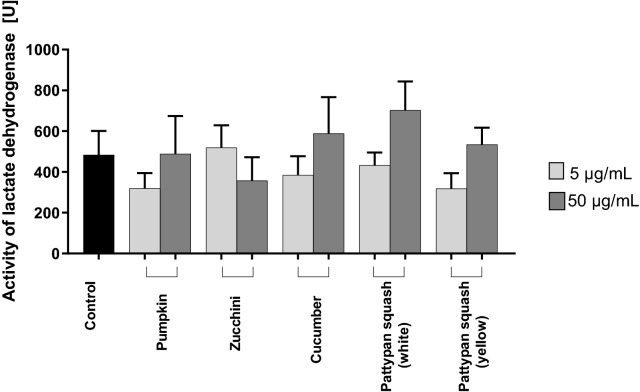
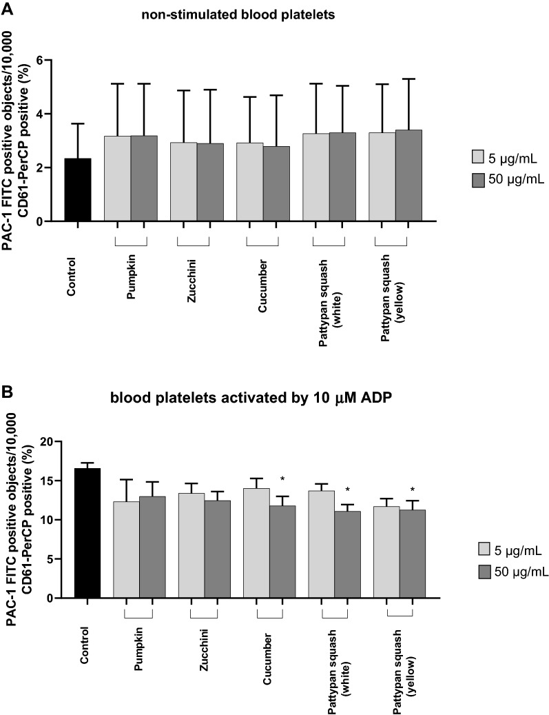
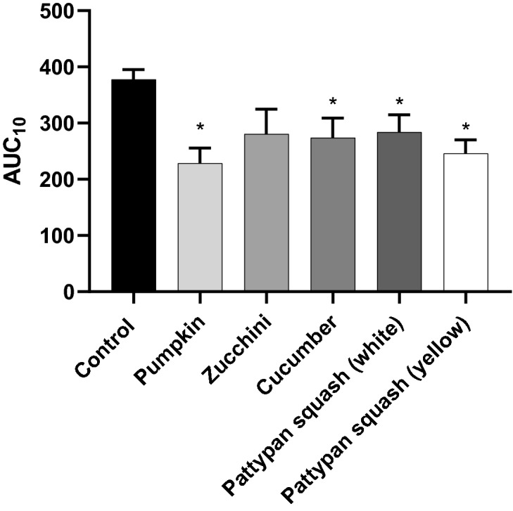
Increased blood platelet activation plays an important role in cardiovascular diseases (CVDs). Recent experiments indicate that certain fruits and vegetables, including onion, garlic, and beetroot, have anti-platelet potential and therefore may reduce the likelihood of CVDs. While vegetables from the Cucuritaceae family are known to exerting beneficial antioxidant and anti-inflammatory effects, their effects on blood platelet activation are poorly understood. Therefore, the aim of the present study was to determine the effect on platelet adhesion of preparations from selected cucurbits: pumpkin (Cucurbita pepo; fruit without seeds), zucchini (Cucurbita pepo convar. giromontina; fruit with seeds), cucumber (Cucumis sativus; fruit with seeds), white pattypan squash (Cucurbita pepo var. patisoniana; fruit without seeds) and yellow pattypan squash (Cucurbita pepo var. patisoniana, fruit without seeds). It also evaluates the activity of these preparations on enzymatic lipid peroxidation in thrombin-activated washed blood platelets by TBARS assay. The study also determines the anti-platelet properties of these five cucurbit preparations in whole blood by flow cytometry and with the total thrombus-formation analysis system (T-TAS) and evaluates the cytotoxicity of the tested preparations against platelets based on LDH activity. The results indicate that the yellow Cucurbita pepo var. patisoniana preparation demonstrated stronger anti-platelet properties than the other tested preparations, reducing the adhesion of thrombin-activated platelets to collagen/fibrinogen, and inhibiting arachidonic acid metabolism and GPIIb/IIIa expression on 10 µM ADP-activated platelets. None of the preparations was found to cause platelet lysis. Our findings provide new information on the anti-platelet activity of the tested cucurbit preparations and their potential for treating CVDs associated with platelet hyperactivity.


Conflict of interest statement

The authors declare no competing interests.

Figures

Figure 1
Figure 1
Effects of the five cucurbit vegetable preparations (concentrations 5 and 50 µg/mL, incubation time—30 min) on damage to human blood platelets. Results are given as mean ± SD (n = 4); the control sample (platelets without plant preparation). There wasn’t any statistically significant between effect of 5 and 50 µg/mL (p > 0.05). The baseline spectral reading (absorbance) for plant preparations range between 0.009 and 0.0245.
Figure 2
Figure 2
Effects of the five cucurbit vegetable preparations (concentrations 5 and 50 µg/mL, incubation time—30 min) on adhesion of resting platelets to collagen (A) or thrombin (final concentration 0.2 U/mL)—activated platelets (B). In the graphs, the adhesion is expressed as a percentage of the control sample (platelets without plant preparation). Results are given as mean ± SD (n = 5). Kruskal–Wallis test: *p < 0.05, **p < 0.01, compared with control (i.e. not treated with plant preparation). There wasn’t any statistically significant between effect of 5 and 50 µg/mL (p > 0.05). The baseline spectral reading (absorbance) for plant preparations range between 0.00075 and 0.0095.
Figure 3
Figure 3
Effects of the five cucurbit vegetable preparations (concentrations 5 and 50 µg/mL, incubation time—30 min) on adhesion to fibrinogen and thrombin (final concentration 0.2 U/mL)—activated platelets (A) or ADP (final concentration 30 µM)—activated platelets (B). In the graphs, the adhesion is expressed as a percentage of the control sample (platelets without plant preparation). Results are given as mean ± SD (n = 5). Kruskal–Wallis test: *p < 0.05, **p < 0.01, ***p < 0.001, compared with control (i.e. not treated with plant preparation). There wasn’t any statistically significant between effect of 5 and 50 µg/mL. There wasn’t any statistically significant between effect of 5 and 50 µg/mL (p > 0.05). The baseline spectral reading (absorbance) for plant preparations range between 0.0025 and 0.0055.
Figure 4
Figure 4
Effects of the five cucurbit vegetable preparations (concentrations 5 and 50 µg/mL) on lipid peroxidation in blood platelets activated by 5 U/mL thrombin (pre-incubation time with plant preparation—25 min; incubation time with thrombin—5 min). Results are given as mean ± SD (n = 5). Control negative refers to platelets not treated with thrombin, and control positive to platelets treated with thrombin. Kruskal–Wallis test: *p < 0.05, **p < 0.01. There wasn’t any statistically significant between effect of 5 and 50 µg/mL (p > 0.05). The baseline spectral reading (absorbance) for plant preparations range between 0.00065 and 0.0035.
Figure 5
Figure 5
Effects of the five cucurbit vegetable preparations (concentrations 5 and 50 µg/mL, incubation time—30 min) on the expression of the active form of GPIIb/IIIa on resting (A) or agonist-stimulated blood platelets: 10 µM ADP (B), 20 µM ADP (C) and 10 µg/mL collagen (D) in whole blood samples. Additionally effects of three selected preparations (cucumber, pattypan squash white and yellow; 50 µg/mL; 30 min) on the expression of the active form of GPIIb/IIIa in platelets stimulated by 10 µM ADP in whole blood samples (E). This figure demonstrates selected diagrams (E). The blood platelets were distinguished based on the expression of CD61. For each sample, 10,000 CD61-positive objects (blood platelets) were acquired. For the assessment of GPIIb/IIIa expression, samples were labeled with fluorescently conjugated monoclonal antibody PAC-1/FITC. Results are shown as the percentage of platelets binding PAC-1/FITC. Data represent the mean ± SD of six healthy volunteers (each experiment performed in triplicate). *p < 0.05 (vs. control platelets). There wasn’t any statistically significant between effect of 5 and 50 µg/mL (p > 0.05).
Figure 5
Figure 5
Effects of the five cucurbit vegetable preparations (concentrations 5 and 50 µg/mL, incubation time—30 min) on the expression of the active form of GPIIb/IIIa on resting (A) or agonist-stimulated blood platelets: 10 µM ADP (B), 20 µM ADP (C) and 10 µg/mL collagen (D) in whole blood samples. Additionally effects of three selected preparations (cucumber, pattypan squash white and yellow; 50 µg/mL; 30 min) on the expression of the active form of GPIIb/IIIa in platelets stimulated by 10 µM ADP in whole blood samples (E). This figure demonstrates selected diagrams (E). The blood platelets were distinguished based on the expression of CD61. For each sample, 10,000 CD61-positive objects (blood platelets) were acquired. For the assessment of GPIIb/IIIa expression, samples were labeled with fluorescently conjugated monoclonal antibody PAC-1/FITC. Results are shown as the percentage of platelets binding PAC-1/FITC. Data represent the mean ± SD of six healthy volunteers (each experiment performed in triplicate). *p < 0.05 (vs. control platelets). There wasn’t any statistically significant between effect of 5 and 50 µg/mL (p > 0.05).
Figure 6
Figure 6
Effects of the five cucurbit vegetable preparations (concentrations 5 and 50 µg/mL, incubation time—30 min) on expression of P-selectin on resting (A) or agonist-stimulated blood platelets: 10 µM ADP (B), 20 µM ADP (C) and 10 µg/mL collagen (D) in whole blood samples. The blood platelets were distinguished based on the expression of CD61/PerCP. For each sample, 5000 CD61-positive objects (blood platelets) were acquired. For the assessment of P-selectin expression, samples were labeled with fluorescently conjugated monoclonal antibody CD62P. Results are shown as the percentage of platelets expressing CD62P. Results are given as the mean ± SD of six healthy volunteers (each experiment performed in triplicate). There wasn’t any statistically significant between effect of 5 and 50 µg/mL (p > 0.05).
Figure 6
Figure 6
Effects of the five cucurbit vegetable preparations (concentrations 5 and 50 µg/mL, incubation time—30 min) on expression of P-selectin on resting (A) or agonist-stimulated blood platelets: 10 µM ADP (B), 20 µM ADP (C) and 10 µg/mL collagen (D) in whole blood samples. The blood platelets were distinguished based on the expression of CD61/PerCP. For each sample, 5000 CD61-positive objects (blood platelets) were acquired. For the assessment of P-selectin expression, samples were labeled with fluorescently conjugated monoclonal antibody CD62P. Results are shown as the percentage of platelets expressing CD62P. Results are given as the mean ± SD of six healthy volunteers (each experiment performed in triplicate). There wasn’t any statistically significant between effect of 5 and 50 µg/mL (p > 0.05).
Figure 7
Figure 7
Effects of the five cucurbit vegetable preparations (concentration—50 µg/mL, incubation time—30 min) on the T-TAS using the PL-chip in whole blood samples. Whole blood samples were analyzed by the T-TAS at the shear rates of 1000 s−1 on the PL-chips. The area under the curve (AUC10) in PL are shown as closed circles. Data represent the mean ± SD of eight healthy volunteers (each experiment performed in triplicate). *p < 0.05 (vs. control blood).

References

    1. Hou Y, et al. Platelets in hemostasis and thrombosis: Novel mechanisms of fibrinogen-independent platelet aggregation and fibronectin-mediated protein wave of hemostasis. J. Biomed. Res. 2015;29:437–444. - PMC - PubMed
    1. Versteeg HH, et al. New fundamentals in hemostasis. Physiol. Rev. 2013;93:327–358. doi: 10.1152/physrev.00016.2011. - DOI - PubMed
    1. Clemetson KJ. Platelets and primary haemostasis. Thromb. Res. 2012;129:220–224. doi: 10.1016/j.thromres.2011.11.036. - DOI - PubMed
    1. Manon-Jensen T, et al. Collagen-mediated hemostasis. J. Thromb. Haemost. 2016;14:438–448. doi: 10.1111/jth.13249. - DOI - PubMed
    1. Yun S-H, et al. Platelet activation: The mechanisms and potential biomarkers. BioMed Res. Int. 2016;9060143:1–5. doi: 10.1155/2016/9060143. - DOI - PMC - PubMed

MeSH terms

Substances