. 2021 Nov 22;4(1):1317.
doi: 10.1038/s42003-021-02835-2.

High-resolution epitope mapping and characterization of SARS-CoV-2 antibodies in large cohorts of subjects with COVID-19

Collaborators, Affiliations

High-resolution epitope mapping and characterization of SARS-CoV-2 antibodies in large cohorts of subjects with COVID-19

Winston A Haynes et al. Commun Biol. .

Abstract

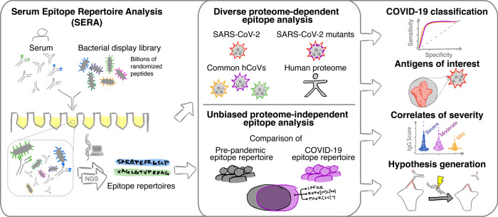
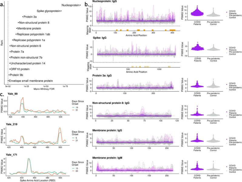
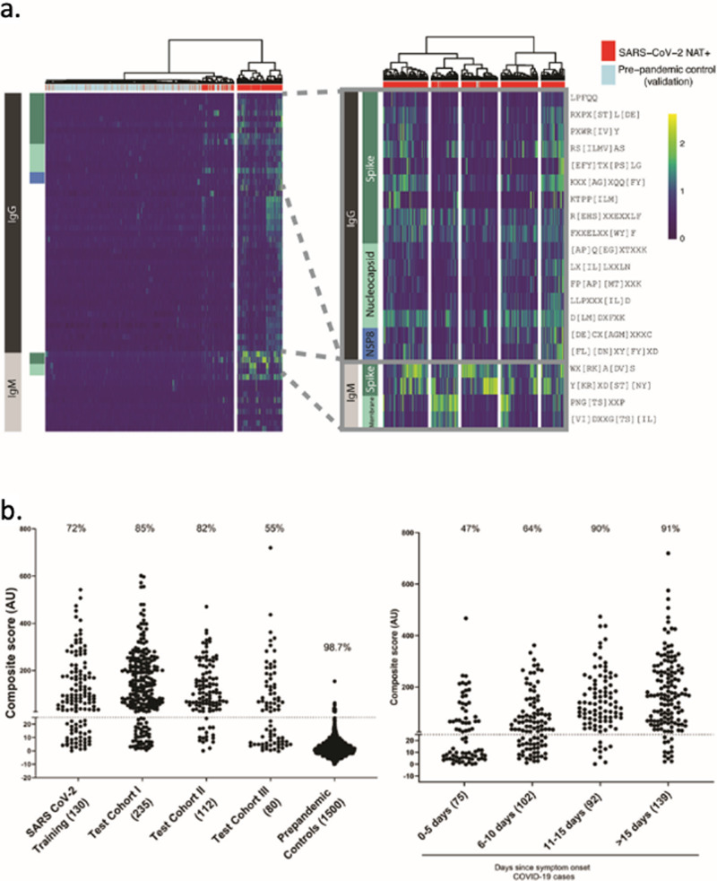
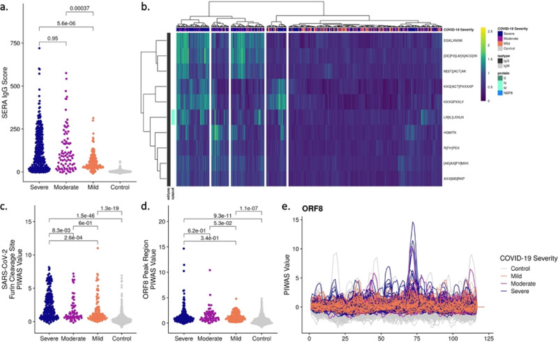
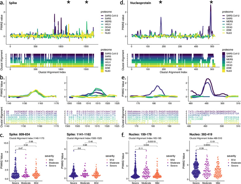
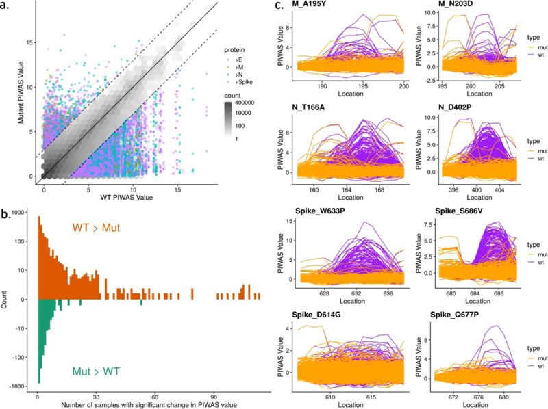
As Severe Acute Respiratory Syndrome Coronavirus 2 (SARS-CoV-2) continues to spread, characterization of its antibody epitopes, emerging strains, related coronaviruses, and even the human proteome in naturally infected patients can guide the development of effective vaccines and therapies. Since traditional epitope identification tools are dependent upon pre-defined peptide sequences, they are not readily adaptable to diverse viral proteomes. The Serum Epitope Repertoire Analysis (SERA) platform leverages a high diversity random bacterial display library to identify proteome-independent epitope binding specificities which are then analyzed in the context of organisms of interest. When evaluating immune response in the context of SARS-CoV-2, we identify dominant epitope regions and motifs which demonstrate potential to classify mild from severe disease and relate to neutralization activity. We highlight SARS-CoV-2 epitopes that are cross-reactive with other coronaviruses and demonstrate decreased epitope signal for mutant SARS-CoV-2 strains. Collectively, the evolution of SARS-CoV-2 mutants towards reduced antibody response highlight the importance of data-driven development of the vaccines and therapies to treat COVID-19.


Conflict of interest statement

The authors declare the following competing interests: employment and personal financial interests including stock options at Serimmune, In: W.A.H., K.K., J.B., E.B.J., P.S.D., A.D., M.J., G.J., B.M., J.R., J.R.S., R.W., M.Z., J.C.S. A.K. reports grant support from Regeneron and Merck for COVID-19 studies, contract and consulting fees from Tata for diagnostics development, honoraria for a COVID-19 lecture from Harvard University, and a research gift from Serimmune. Serimmune has also submitted the following US Patent Application 63/114,939 SARS-CoV-2 Serum Antibody Profiling SUI-009PR4. The remaining authors declare no competing interests.

Figures

Fig. 1
Fig. 1. The Serum Epitope Repertoire Analysis (SERA) platform enables high-resolution mapping of SARS-CoV-2 antibody repertoires.
The SERA assay results in a set of ~1 million unique peptides, the “epitope repertoire”, for each individual. Repertoires were deposited in a database and compared with pre-pandemic controls to identify conserved epitopes in SARS-CoV-2 using proteome-dependent and -independent bioinformatic methods. SERA enables analysis of COVID-19 repertoires against any proteome including mutant SARS-CoV-2 strains, human common coronaviruses and the human proteome for discovery of potential autoantigens. The identified epitope signatures can be used to build diagnostic classifiers, to identify correlates of disease severity, and to develop hypotheses based on cases with specific symptoms and/or disease course (neurological, GI, cardio e.g.).
Fig. 2
Fig. 2. Bioinformatic analysis of SERA antibody repertoires identifies the antigens and epitopes involved in the SARS-CoV-2 immune response.
a PIWAS statistical ranking of kmer enrichments across the SARS-CoV-2 proteome using the Mann–Whitney false-discovery rate (FDR). Multiple antigens in addition to spike and nucleoprotein showed significant enrichment for one or more epitopes. b PIWAS kmer enrichments from COVID-19 repertoires versus pre-pandemic controls across statistically significant antigens. PIWAS values = number of standard deviations above the mean of 1500 pre-pandemic controls. PIWAS tiling (left): Dark purple = average COVID-19 patient signal, light purple = 95th quantile band for COVID-19 patient signal, dark gray = average pre-pandemic control patient signal, light gray = 95th quantile band for pre-pandemic patient signal. PIWAS distribution (right): One point per patient of maximum PIWAS value across each protein. IMUNE motifs largely mapped to the same prominent epitopes that were identified by PIWAS. Epitopes on spike and nucleoprotein discovered by IMUNE are shown below each antigen (orange bars). All antigens were found to be statistically different from controls as shown in a. c Longitudinal samples from individual subjects enabled identification of RBD-specific signals that emerged over time but were not conserved across COVID-19 patients.
Fig. 3
Fig. 3. IMUNE-based discovery of IgG and IgM motifs in the SARS-CoV-2 humoral immune response.
a Heatmap of IgG and IgM motif log-enrichment values for 579 COVID-19 samples and 1500 pre-pandemic controls. Inset highlights motifs with linear epitope maps to SARS-CoV-2. b Sensitivity and specificity of the SARS-CoV-2 IgG/IgM diagnostic classifier in NAT + subjects and pre-pandemic controls. Z-scores for each motif were summed to generate an IgG/IgM composite score. The maximum value for the IgG or IgM for each sample i shown. Samples above a cutoff of 25 are classified as positive. The sensitivity or specificity of the SERA panels for all COVID-19 cohorts and controls is shown above each column.
Fig. 4
Fig. 4. Significantly different epitope signals are observed in mild, moderate, and severe cases of COVID-19.
a Comparison of SERA total IgG motif panel scores for severe (n = 447), moderate (n = 134), and mild (n = 170) cases based on motifs used in the diagnostic panel (Fig. 3). Colors indicate disease severity. b Severe, moderate, and mild cases of SARS-CoV-2 are clustered based on log-enrichment of the top 10 motifs identified by a t-test comparison of severe and mild patients. c Distribution of PIWAS values at SARS-CoV-2 furin cleavage site for severe, moderate, and mild cases. d Distribution of PIWAS values for the peak epitope in ORF8 for severe, moderate, and mild cases. e PIWAS tiling of individual samples on the entire ORF8 sequence. All p-values were calculated using outlier sum statistical test.
Fig. 5
Fig. 5. Cross-reactivity analysis across coronaviruses reveals shared epitopes and epitopes specific to SARS-CoV-2.
PIWAS was performed using COVID-19 samples against various coronavirus proteomes including SARS-CoV-2, SARS, MERS, and the common hCoVs HKU1, OC43, 229E, and NL63. PIWAS tilings for a spike and d nucleoprotein revealed regions of cross-reactivity as well as epitopes only observed against SARS-CoV-2. Clustal multiple sequence alignments were performed and visualized below to depict sequence similarity and divergence. Distinct epitopes from b spike and e nucleoprotein showcase PIWAS values across the coronaviruses with corresponding clustal alignment sequences below. Epitope locations are denoted with asterisks in a and d. Distribution of PIWAS values at epitopes from spike c and nucleoprotein f for severe (n = 447), moderate (n = 134), and mild (n = 170) cases, with p-values from Wilcoxon-rank sum test.
Fig. 6
Fig. 6. Mutations to SARS-CoV-2 are biased towards decreasing immune epitope response.
21,127 distinct amino acid mutations in spike glycoprotein, nucleoprotein, envelope protein, and membrane protein in SARS-CoV-2 strains were identified from sequencing data of 96,437 genomes from GISAID. a For each mutation, the PIWAS value of the wild-type (WT) sequence was compared to the PIWAS value for the mutated strain (mut). b Mutations conferring a significant PIWAS value change (|PIWASWT-PIWASmut | > 3) for each COVID-19 sample were identified. For each mutation, the number of patients with a significant difference was counted. c Exemplary mutations that yielded a decrease in PIWAS values are shown for membrane protein (top row), nucleoprotein (middle row), and spike (bottom row). No significant immune signal is seen at location 614 of spike, for either the wild-type or the D614G variant. The Q677P mutation in spike resulted in a loss of signal at that epitope.

References

    1. Nishiura H, et al. Estimation of the asymptomatic ratio of novel coronavirus infections (COVID-19) Int. J. Infect. Dis. 2020;94:154–155. doi: 10.1016/j.ijid.2020.03.020. - DOI - PMC - PubMed
    1. Mizumoto, K., Kagaya, K., Zarebski, A. & Chowell, G. Estimating the asymptomatic proportion of coronavirus disease 2019 (COVID-19) cases on board the Diamond Princess cruise ship, Yokohama, Japan, 2020. Eurosurveillance25, 2000180 (2020). - PMC - PubMed
    1. Atyeo C, et al. Distinct early serological signatures track with SARS-CoV-2 survival. Immunity. 2020;53:524–532.e4. doi: 10.1016/j.immuni.2020.07.020. - DOI - PMC - PubMed
    1. Amrun, S. N. et al. Linear B-cell epitopes in the spike and nucleocapsid proteins as markers of SARS-CoV-2 exposure and disease severity. EBioMedicine58, 102911 (2020). - PMC - PubMed
    1. Mathew, D. et al. Deep immune profiling of COVID-19 patients reveals distinct immunotypes with therapeutic implications. Science369 (2020). - PMC - PubMed