

Spatial mapping of protein composition and tissue organization: a primer for multiplexed antibody-based imaging
- PMID: 34811556
- PMCID: PMC9264278
- DOI: 10.1038/s41592-021-01316-y
Spatial mapping of protein composition and tissue organization: a primer for multiplexed antibody-based imaging
Abstract
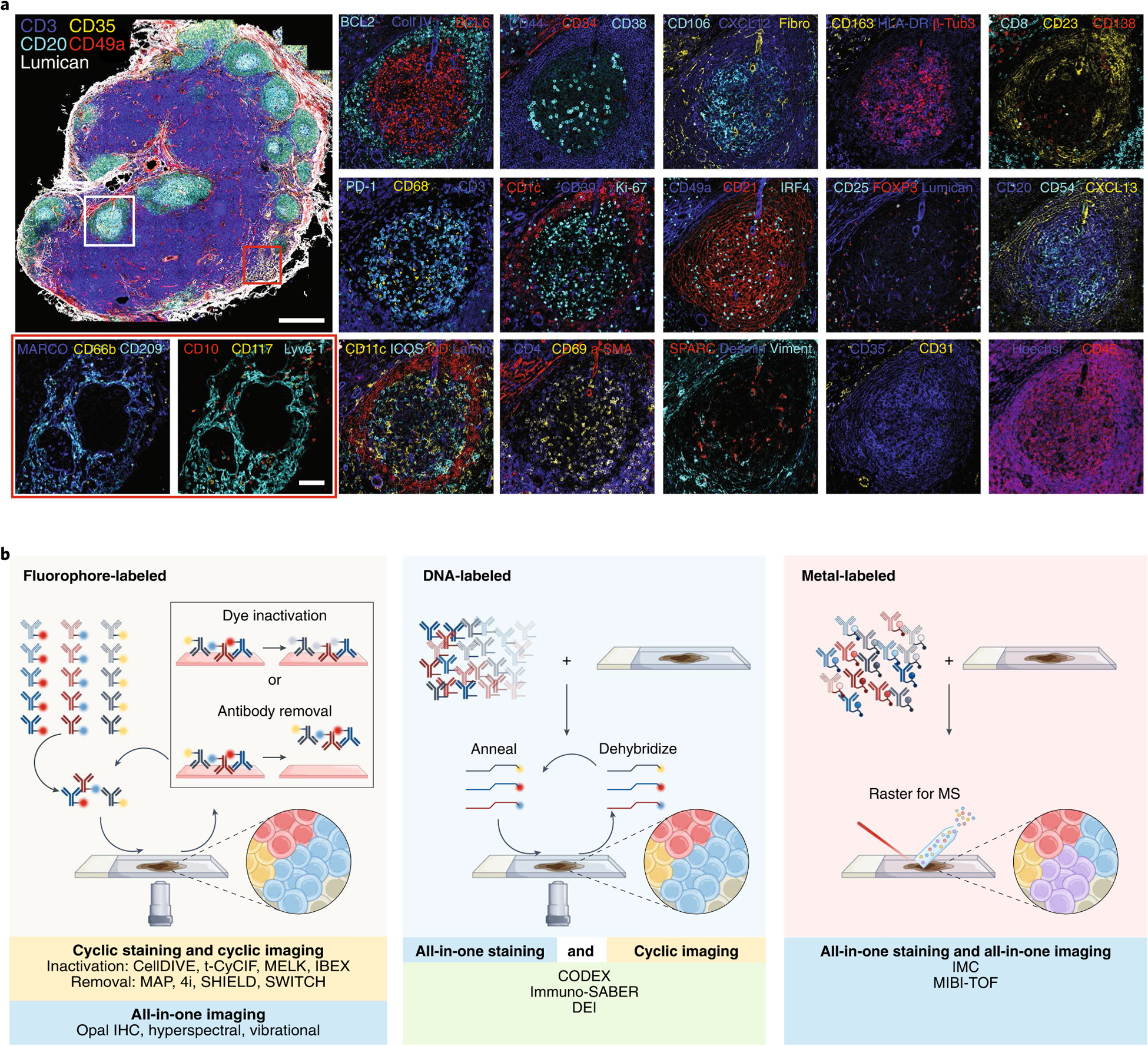
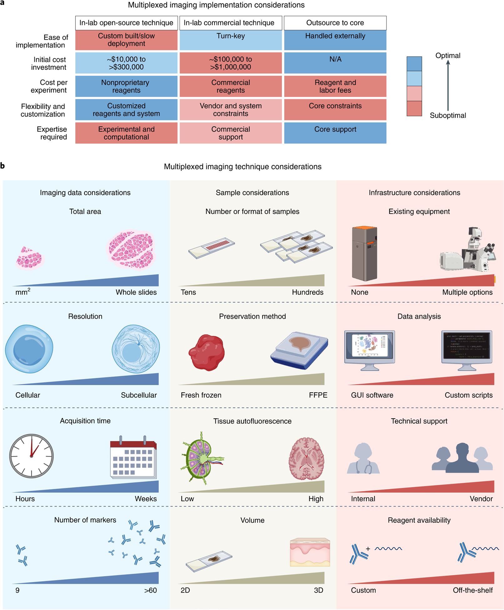
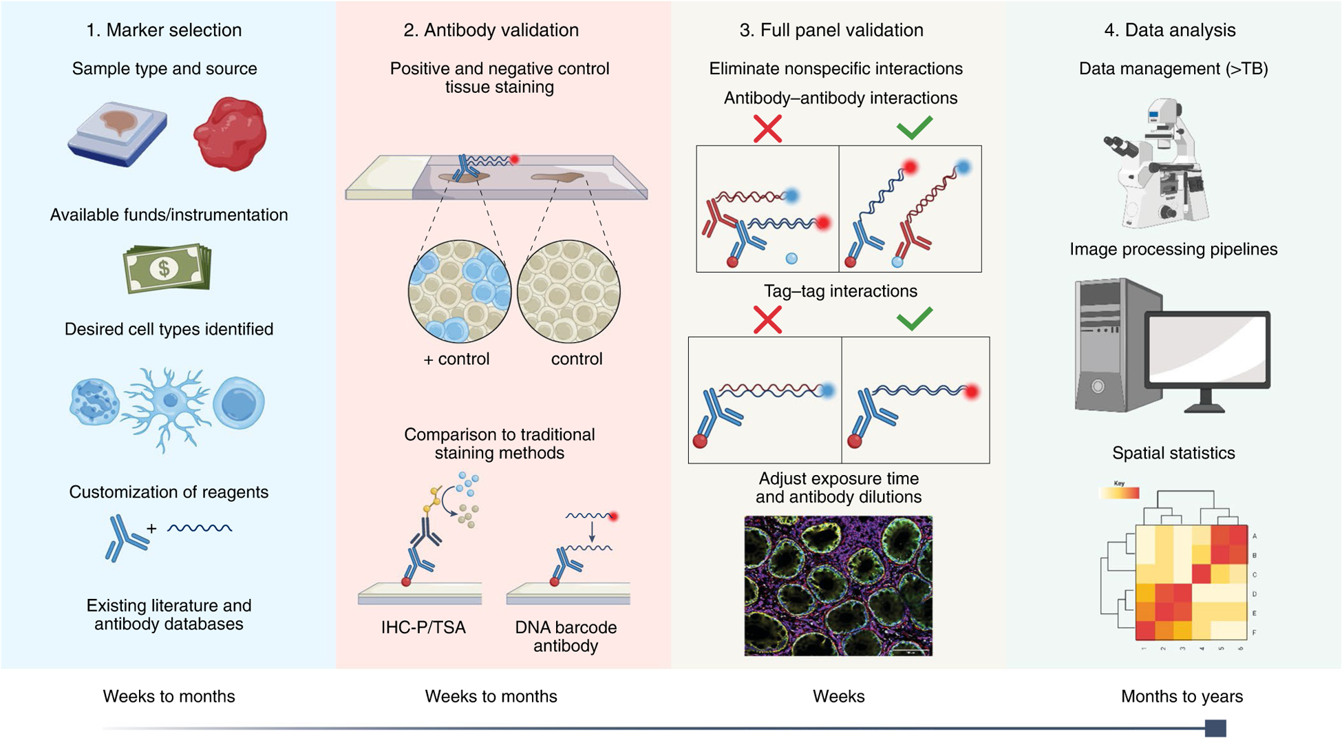
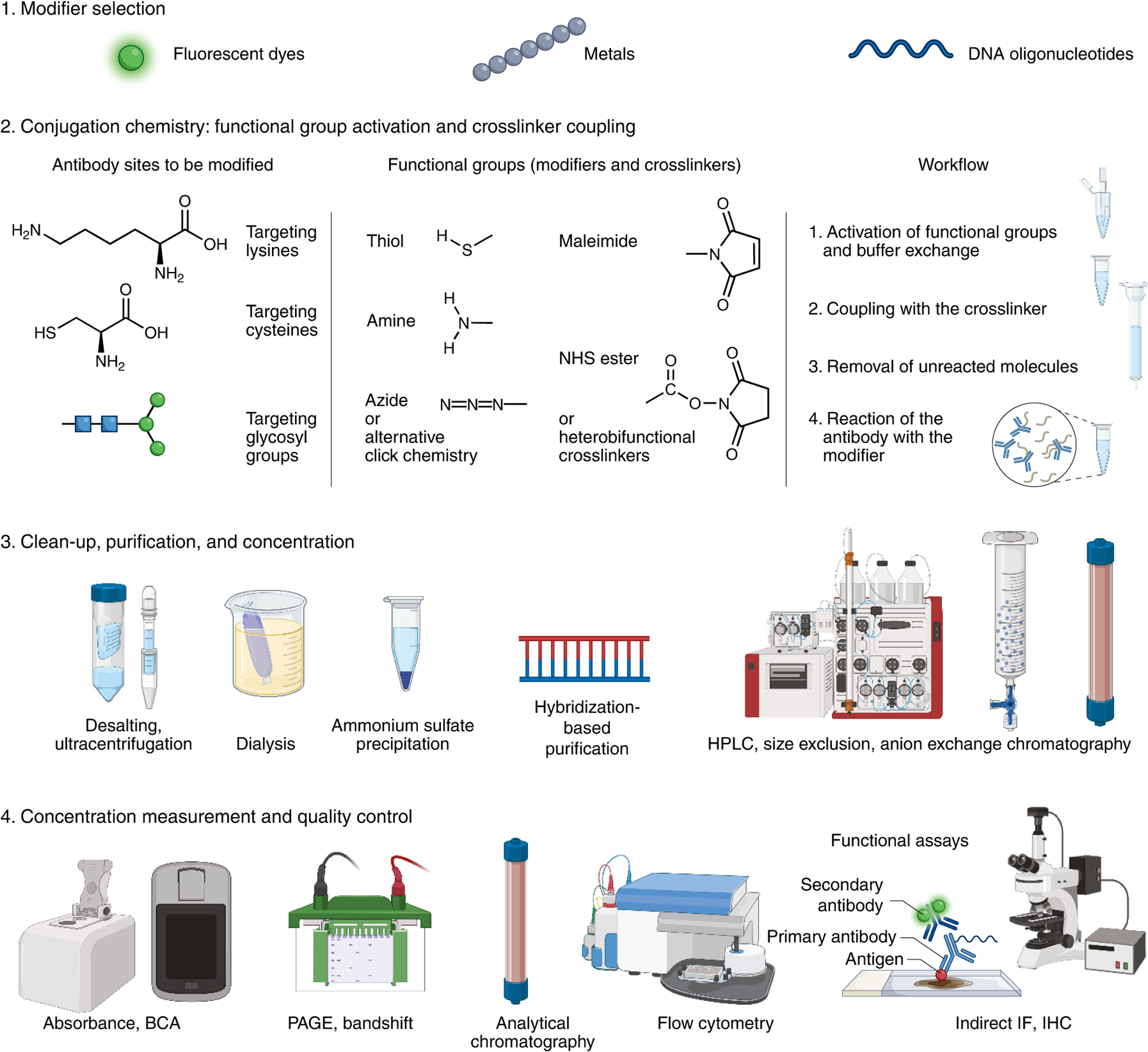
Tissues and organs are composed of distinct cell types that must operate in concert to perform physiological functions. Efforts to create high-dimensional biomarker catalogs of these cells have been largely based on single-cell sequencing approaches, which lack the spatial context required to understand critical cellular communication and correlated structural organization. To probe in situ biology with sufficient depth, several multiplexed protein imaging methods have been recently developed. Though these technologies differ in strategy and mode of immunolabeling and detection tags, they commonly utilize antibodies directed against protein biomarkers to provide detailed spatial and functional maps of complex tissues. As these promising antibody-based multiplexing approaches become more widely adopted, new frameworks and considerations are critical for training future users, generating molecular tools, validating antibody panels, and harmonizing datasets. In this Perspective, we provide essential resources, key considerations for obtaining robust and reproducible imaging data, and specialized knowledge from domain experts and technology developers.
© 2021. Springer Nature America, Inc.
Conflict of interest statement
Competing interests
A. E. W. is an employee and shareholder of Abcam plc. J. F. is an employee of Cell Signaling Technologies. J. C. is an employee and shareholder of BioLegend. J. H. is an employee of Bio-Techne. E. S. is an employee of Thermo Scientific. E. M. and A. S. are current or past employees of GE Research. K. C. is an inventor for patent applications covering some technologies described in this paper and a cofounder of LifeCanvas Technologies. G. P. N. is inventor on a US patent, covering some technologies described in this paper, has equity in and/or is a member of the scientific advisory board of Akoya Biosciences. S. K. S. is an inventor for patent applications related to some of the methods described here.
Figures
References
-
- Stack EC, Wang C, Roman KA & Hoyt CC Multiplexed immunohistochemistry, imaging, and quantitation: A review, with an assessment of Tyramide signal amplification, multispectral imaging and multiplex analysis. Methods 70, 46–58 (2014). - PubMed
-
- Thul PJ et al. A subcellular map of the human proteome. Science 356, eaal3321 (2017). - PubMed
-
- Gawad C, Koh W & Quake SR Single-cell genome sequencing: current state of the science. Nat. Rev. Genet 17, 175–188 (2016). - PubMed
-
- Massonnet P & Heeren RMA A concise tutorial review of TOF-SIMS based molecular and cellular imaging. J. Anal. Spectrom 34, 2217–2228 (2019).
Publication types
MeSH terms
Substances
Grants and funding
- UH3 CA246635/CA/NCI NIH HHS/United States
- UH3 CA255133/CA/NCI NIH HHS/United States
- U54 DK134302/DK/NIDDK NIH HHS/United States
- U54 DK120058/DK/NIDDK NIH HHS/United States
- UG3 HL145600/HL/NHLBI NIH HHS/United States
- R01 AI145992/AI/NIAID NIH HHS/United States
- U54 AR081775/AR/NIAMS NIH HHS/United States
- U54 HG010426/HG/NHGRI NIH HHS/United States
- T32 ES007028/ES/NIEHS NIH HHS/United States
- P30 DK020593/DK/NIDDK NIH HHS/United States
- UH3 CA246594/CA/NCI NIH HHS/United States
- T32 CA196585/CA/NCI NIH HHS/United States
- R01 AI138581/AI/NIAID NIH HHS/United States
- U54 EY032442/EY/NEI NIH HHS/United States
- UH3 CA246633/CA/NCI NIH HHS/United States
LinkOut - more resources
Full Text Sources
Other Literature Sources
Research Materials
