Review
. 2021 Nov;19(11):1571-1580.
doi: 10.1111/ddg.14582.

Hide-and-seek: Neurotropic squamous cell carcinoma of the periorbital region - a series of five cases and review of the literature

Affiliations
Review

Hide-and-seek: Neurotropic squamous cell carcinoma of the periorbital region - a series of five cases and review of the literature

Evi M Morandi et al. J Dtsch Dermatol Ges. 2021 Nov.

Abstract

Squamous cell carcinoma is the second most common malignancy of the skin after basal cell carcinoma and mainly found in sun-exposed areas such as the face. This mostly locally destructive malignancy may show invasive growth and insidious mechanisms of dissemination such as perineural invasion. Periorbital squamous cell carcinoma is associated with perineural invasion in up to 14 % of cases. Specifically in this region, the proximity to cranial nerves and therefore the associated risk of progression to the central nervous system are associated with poor prognosis. The clinically concealed character of this entity often leads to a delay in diagnosis and consequently makes complete resection and reconstruction demanding. Careful clinical evaluation often hints at perineural invasion before obtaining histology. Aside from presenting five challenging cases, this work analyzes risk factors, clinical as well as histological features, and treatment options for periorbital squamous cell carcinoma with perineural invasion.


Conflict of interest statement

None.

Figures

Figure 1
Figure 1
Case 1: Six months after complete resection of squamous cell carcinoma with perineural invasion of the left frontal region. Recurrence was diagnosed by biopsy. Computed tomography scans were performed because of suspected invasion of bone; neoplastic growth along the supraorbital nerve was found (arrow) (a). Resection included frontal bone. Reconstruction was performed with Palacos bone cement followed by free‐flap coverage (b).
Figure 2
Figure 2
Case 4: The patient was referred with a rapidly growing neoplastic mass in the left temporal area. After four operations, free margins were achieved and the defect was covered with a full‐thickness skin graft and a local flap. However, after five months the patient presented with painful pressure sensation in the left orbit accompanied by double vision (a). MRI scans (1.5 T; axial T1‐weigthed VIBE DIXON sequence after intravenous application of gadolinium‐based contrast agent) revealed perineural and direct invasion of the orbit, with obliteration of the inferior orbital fissure (dotted line) (b) and of the pterygopalatine fossa (arrow) (c).
Figure 3
Figure 3
Case 5: Five months after primary resection; the patient was referred to our outpatient clinic. The locally persisting process was mistaken for dacryocystitis on initial presentation (a). One year after excision (1.5 T MRI with axial T1‐weigthed TSE DIXON sequence after intravenous application of gadolinium‐based contrast agent): Recurrence (dotted line) with intraorbital perineural invasion was diagnosed along the supratrochlear and infraorbital nerve extending to the optic nerve and eventually reaching the optic chiasma (b).
Figure 4
Figure 4
Histology of Case 1: Perineural invasion (small arrows) along hyperplastic nerve (H&E stain) (a). Double stain with reddish S100 protein for Schwann cells of nerve and brown nuclear P40 staining of scirrhous squamous cell carcinoma (small arrows); positive p40 stain by nuclei of perineural cells (thick arrow) (b). The star marks corresponding areas of Indian filing invasion of scirrhous squamous cell carcinoma in fibrosclerotic background (a, b). Histology of Case 5: Subtle perineural spread (small arrows) of nuclei from scirrhous squamous cell carcinoma (hematoxylin‐eosin stain) (c). Double stain with S100 protein for Schwann cells and nuclear staining with p40 for scirrhous squamous cell carcinoma (small arrow). Positive p40 stain by nuclei of perineural cells (thick arrow) (d). Histology of Case 4: Intraorbital perimuscular extension: Perimysial spread of scirrhous squamous cell carcinoma between fascicles of striated muscle tissue; note multiple mitoses (thick arrows) in addition to dense cellularity, nuclear pleomorphism and hyperchromasia (e). Scale bar is 300 μm.
Figure 5
Figure 5
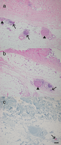
Hematoxylin‐eosin (HE) stain showing two hyperplastic nerves (arrow heads) at the border of dermis to subcutaneous layer; lymph follicle‐like aggregation of lymphocytes (arrows) in close proximity (a). Serial section of HE stains with loss of left nerve and inflammatory infiltrate; to the right, area with lymph follicle‐like aggregation of lymphocytes again in close proximity to hyperplastic nerve (arrowhead). Tiny focus of dense, hyperchromatic nuclei in right lower corner (arrow) (b). Serial section showing reactivity of neoplastic cells with AE1/AE3 stain (arrow) of this scirrhous SCC. The separation in comparison to the previous cutting level indicates infiltration of scirrhous SCC. Close perineural contact is not present in these cutting levels (c). Scale bar is 500 μm.
Figure 6
Figure 6
Depiction of the periorbital region.
Figure 7
Figure 7
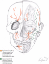
Anatomical depiction of nerves in the facial and periorbital region. The trigeminal nerve (cranial nerve V) with its three terminal branches, the ophthalmic (V1), the maxillary (V2) and the mandibular (V3) nerve, is shown in orange. The facial nerve (cranial nerve VII) is shown in green. The supratrochlear nerve (1) was involved in our clinical Case 3. The supraorbital nerve (2) also showed neoplastic invasion in Case 3. The infraorbital nerve (3) was affected in Case 1, Case 2 and Case 4. The auriculotemporal nerve (4) was involved in Case 5. Buccal branches of the facial nerve (c) were also affected in Case 2.

References

    1. Leiter U, Garbe C. Epidemiology of melanoma and nonmelanoma skin cancer – the role of sunlight. Adv Exp Med Biol 2008; 624: 89–103. - PubMed
    1. Leiter U, Heppt MV, Steeb T et al. S3 guideline for actinic keratosis and cutaneous squamous cell carcinoma (cSCC) – short version, part 2: epidemiology, surgical and systemic treatment of cSCC, follow‐up, prevention and occupational disease. J Dtsch Dermatol Ges 2020; 18(4): 400–13. - PubMed
    1. Brantsch KD, Meisner C, Schönfisch B et al. Analysis of risk factors determining prognosis of cutaneous squamous‐cell carcinoma: a prospective study. Lancet Oncol 2008; 9(8): 713–20. - PubMed
    1. Batsakis JG. Nerves and neurotropic carcinomas. Ann Otol Rhinol Laryngol 1985; 94(4 Pt 1): 426–7. - PubMed
    1. Moore CE, Hoyt WF, North JB. Painful ophthalmoplegia following treated squamous carcinoma of the forehead. Orbital apex involvement from centripetal spread via the supraorbital nerve. Med J Aust 1976; 1(18): 657–9. - PubMed