Review
. 2021 Dec 17;49(6):2697-2710.
doi: 10.1042/BST20210672.

Molecular and structural aspects of gasdermin family pores and insights into gasdermin-elicited programmed cell death

Affiliations
Review

Molecular and structural aspects of gasdermin family pores and insights into gasdermin-elicited programmed cell death

Ayesha Zahid et al. Biochem Soc Trans. .

Abstract

Pyroptosis is a highly inflammatory and lytic type of programmed cell death (PCD) commenced by inflammasomes, which sense perturbations in the cytosolic environment. Recently, several ground-breaking studies have linked a family of pore-forming proteins known as gasdermins (GSDMs) to pyroptosis. The human genome encodes six GSDM proteins which have a characteristic feature of forming pores in the plasma membrane resulting in the disruption of cellular homeostasis and subsequent induction of cell death. GSDMs have an N-terminal cytotoxic domain and an auto-inhibitory C-terminal domain linked together through a flexible hinge region whose proteolytic cleavage by various enzymes releases the N-terminal fragment that can insert itself into the inner leaflet of the plasma membrane by binding to acidic lipids leading to pore formation. Emerging studies have disclosed the involvement of GSDMs in various modalities of PCD highlighting their role in diverse cellular and pathological processes. Recently, the cryo-EM structures of the GSDMA3 and GSDMD pores were resolved which have provided valuable insights into the pore formation process of GSDMs. Here, we discuss the current knowledge regarding the role of GSDMs in PCD, structural and molecular aspects of autoinhibition, and pore formation mechanism followed by a summary of functional consequences of gasdermin-induced membrane permeabilization.

Keywords: gasdermin; inflammasome; pore-forming proteins; programmed cell death; pyroptosis.


Conflict of interest statement

The authors declare that there are no competing interests associated with the manuscript.

Figures

Figure 1.
Figure 1.. GSDMs can be activated by diverse range of enzymes leading to the induction of various forms of programmed cell death.
(A) Diverse microbial and cellular stress signals are detected by the sensor proteins of the canonical inflammasomes resulting in the recruitment of adaptor protein ASC and procaspase-1 resulting in inflammasome assembly which yields active caspase-1. Proinflammatory cytokines pro-IL-1β and pro-IL-18 are processed by caspase-1 to release mature IL-1β and IL-18. Caspase-1 can initiate pyroptosis by cleaving GSDMD to generate active GSDMD-NT which forms pores in the plasma membrane by targeting phospholipids resulting in pyroptosis, although other caspase-1 substrates capable of inducing pyroptosis may also exist. IL-1β and IL-18 are presumably released by cell lysis during pyroptosis. (B) The non-canonical inflammasome pathway is triggered by cytosolic Gram-negative bacteria or bacterial LPS in the infected cells resulting in activation of caspase 11 in mice (caspase-4/5 in humans) which cleaves GSDMD initiating pyroptosis. In the first step, the GSDMD pores allow potassium release, resulting in the activation of the NLRP3 inflammasome and IL-1β/IL-18 maturation. In a second step, GSDMD pores cause pyroptosis, thereby driving the release of mature cytokines. (C) Granzymes initiate GSDMB-NT- and GSDME-NT-induced pyroptosis. GzmA or GzmB, which is released from killer cytotoxic lymphocytes, induces GSDMB- or GSDME-dependent pyroptosis in tumor cells, respectively. (D) The intrinsic or extrinsic apoptotic pathway can activate caspase-3 which cleaves GSDME to produce the pyroptotic GSDME-NT, leading to membrane permeabilization and releases of proinflammatory DAMP molecules such as high mobility group box protein 1 (HMGB1), subsequently inducing secondary necrosis in macrophages. ASC, apoptosis-associated speck-like protein containing a CARD; AIM2, absent from melanoma 2; GSDMD, gasdermin D; GSDME, gasdermin E; GSDMB, gasdermin B; GzmA, granzyme A; GzmB, granzyme B; HMGB1, high mobility group box protein 1; IL-1β, interleukin-1β; IL-18, interleukin-18, LPS, lipopolysaccharide; NLRP3, Nod-like receptor (NLR) pyrin domain-containing 3; NLRC4, NLR family CARD domain-containing protein 4.
Figure 2.
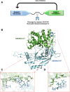
Figure 2.. Two domain architecture and crystal structure of GSDMA3.
(A) The two domain architecture of the GSDM family. Before proteolytic activation, the pore-forming GSDMA3-NT is kept inactive by the auto-inhibitory GSDMA3-CT. (B) X-ray crystal structure of murine GSDMA3 (PDB: 5B5R) is shown as a cartoon model with labeled secondary structure elements. The structure shows that GSDMA3-CT, colored in green, folds back on the functional GSDMA3-NT, colored in light-blue, for autoinhibition. (C,D) The two domain interaction sites I and II are shown. Residues involved in the auto-inhibitory interactions are labeled and shown as sticks.
Figure 3.
Figure 3.. Mechanism of GSDM pore formation.
(A) Crystal structure of autoinhibited GSDMA3 (PDB: 5B5R). (B) Ribbon diagram of GSDMA3-NT domain in the pore conformation. (C) Conformational changes from autoinhibited to membrane-inserted GSDMA3-NT. Extension domains ED1 shown in gray color and ED2 shown in blue color, which transition into the finger-like hairpins HP1 and HP2, respectively. (D) The cryo-EM structure of the 27-subunit murine GSDMA3 pore (PDB: 6CB8). In addition to the 108-strand β-barrel that inserts into the membrane, each subunit contributes a basic α1 helix (orange) that interacts with acidic lipids and a globular domain (green) above the transmembrane (TM) region (adapted from [64]). (E) Ribbon diagram and dimensions of the 33-subunit human GSDMD pore structure fitted into its cryo-EM density map. The 33-fold pore comprises a large transmembrane β-barrel and a globular domain on the cytosolic side of the pore (adapted from [67]).

References

    1. Hu, Z., Zhou, Q., Zhang, C., Fan, S., Cheng, W., Zhao, Y.et al. (2015) Structural and biochemical basis for induced self-propagation of NLRC4. Science 350, 399–404 10.1126/science.aac5489 - DOI - PubMed
    1. Tenthorey, J.L., Haloupek, N., López-Blanco, J.R., Grob, P., Adamson, E., Hartenian, E.et al. (2017) The structural basis of flagellin detection by NAIP5: a strategy to limit pathogen immune evasion. Science 358, 888–893 10.1126/science.aao1140 - DOI - PMC - PubMed
    1. Zhang, L., Chen, S., Ruan, J., Wu, J., Tong, A.B., Yin, Q.et al. (2015) Cryo-EM structure of the activated NAIP2-NLRC4 inflammasome reveals nucleated polymerization. Science 350, 404–409 10.1126/science.aac5789 - DOI - PMC - PubMed
    1. Shen, C., Sharif, H., Xia, S. and Wu, H. (2019) Structural and mechanistic elucidation of inflammasome signaling by cryo-EM. Curr. Opin. Struct. Biol. 58, 18–25 10.1016/j.sbi.2019.03.033 - DOI - PMC - PubMed
    1. Li, Y., Huang, Y., Cao, X., Yin, X., Jin, X., Liu, S.et al. (2018) Functional and structural characterization of zebrafish ASC. FEBS J. 285, 2691–2707 10.1111/febs.14514 - DOI - PMC - PubMed

Publication types

Substances