. 2022 Feb;38(2):287-294.
doi: 10.1007/s00381-021-05408-0. Epub 2021 Nov 23.

Comparative anatomy of dissected optic lobes, optic ventricles, midbrain tectum, collicular ventricles, and aqueduct: evolutionary modifications as potential explanation for non-tumoral aqueductal anomalies in humans

Affiliations

Comparative anatomy of dissected optic lobes, optic ventricles, midbrain tectum, collicular ventricles, and aqueduct: evolutionary modifications as potential explanation for non-tumoral aqueductal anomalies in humans

E Leon Kier et al. Childs Nerv Syst. 2022 Feb.

Abstract

Purpose: An extensive literature has postulated multiple etiologies for aqueductal stenosis. No publications were found, discussing that evolutionary modifications might explain aqueductal anomalies. This study's objectives were to review the evolutionary modifications of vertebrates' tectum structures that might explain human aqueduct anomalies. Undertaking vertebrate comparative study is currently not feasible in view of limitations in obtaining vertebrate material. Thus, vertebrate material collected, injected, dissected, and radiographed in the early 1970s was analyzed, focusing on the aqueduct and components of the midbrain tectum.

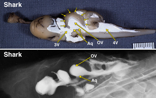
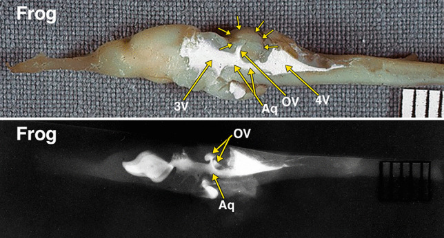
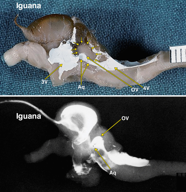
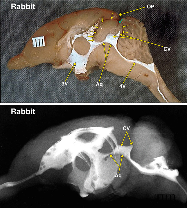
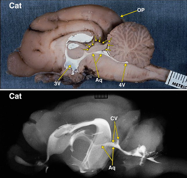
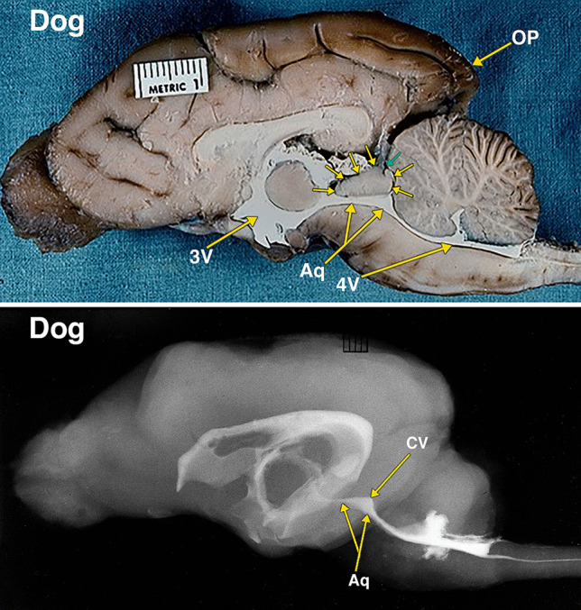
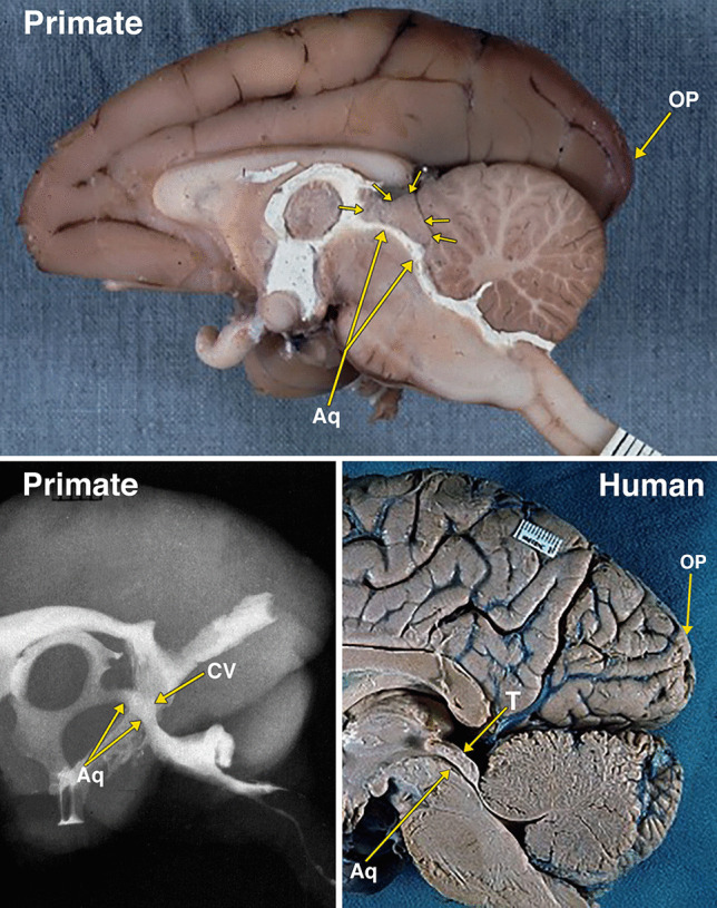
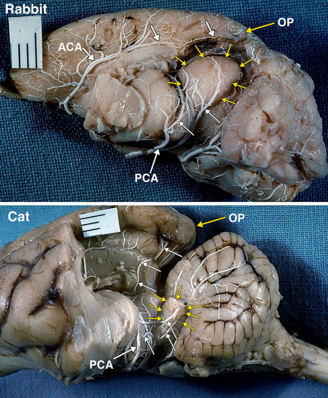
Methods: Photographs of brain dissections and radiographs of the cerebral ventricles and arteries of adult shark, frog, iguana, rabbit, cat, dog, and primate specimens, containing a barium-gelatin radiopaque compound, were analyzed focusing on the aqueduct, the optic ventricles, the quadrigeminal plate, and collicular ventricles. The anatomic information provided by the dissections and radiographs is not reproducible by any other radiopaque contrast currently available.

Results: Dissected and radiographed cerebral ventricular and arterial systems of the vertebrates demonstrated midbrain tectum changes, including relative size modifications of the mammalian components of the tectum, simultaneously with the enlargement of the occipital lobe. There is a transformation of pre-mammalian optic ventricles to what appear to be collicular ventricles in mammals, as the aqueduct and collicular ventricle form a continuous cavity.

Conclusions: The mammalian tectum undergoes an evolutionary cephalization process consisting of relative size changes of the midbrain tectum structures. This is associated with enlargement of the occipital lobe, as part of overall neocortical expansion. Potentially, aqueductal anomalies could be explained by evolutionary modifications.

Keywords: Anatomy Comparative; Cerebral aqueduct; Dissection; Mammals; Nonmammalian; Optic lobe; Tectum Mesencephali.


Conflict of interest statement

The authors declared no potential conflicts of interest with respect to the research, authorship, and/or publication of this article.

Figures

Fig. 1
Fig. 1
Dissected brain of a shark with radiopaque barium-gelatin-contrast filled cerebral ventricles. The midsagittal dissection (Fig. 1) demonstrates a relatively large, thin-walled optic lobe, outlined by multiple small arrows, that includes a large optic ventricle (OV). The midbrain ventricle (Aq) and optic ventricles form a large continuous cavity. The comparative anatomy literature uses the term aqueduct for the midbrain ventricle in all vertebrates with a cerebral ventricular system. The lateral radiograph of the entire dissected brain demonstrates both superimposed optic ventricles (OV) and the aqueduct (Aq). Additional abbreviations: 3 V, 3d ventricle; 4 V, 4th ventricle. The distance between 2 black lines on the ruler in the specimen photograph is 1 mm
Fig. 2
Fig. 2
Dissected brain of a frog with radiopaque contrast filled cerebral ventricles. The midsagittal dissection (Fig. 2) demonstrates a prominent optic lobe, outlined by multiple small arrows, and includes only a small central segment of the optic ventricles (OV) extending superiorly from the aqueduct (Aq). The remainder of the more laterally positioned optic ventricles is visualized in the radiograph. The lateral radiograph of the entire dissected brain demonstrates both optic ventricles (OV) and the aqueduct (Aq). The constricted communication between the dorsolateral segment of the optic ventricles and medial segment of the opticventricle (aqueduct) is likely the result of compressed from below by the torus semicircularis [28]. Additional abbreviations: 3 V, 3d ventricle; 4 V, 4th ventricle. The distance between 2 black lines on the ruler in the specimen photograph is 1 mm
Fig. 3
Fig. 3
Dissected brain of an iguana with radiopaque contrast filled cerebral ventricles. The midsagittal dissection (Fig. 3) demonstrates a relatively large optic lobe, outlined by multiple small arrows, that includes an optic ventricle (OV). The midbrain ventricle (Aq) and optic ventricles form a large continuous cavity. The lateral radiograph (Fig. 3B) of the entire dissected brain demonstrates the mostly superimposed optic ventricles (OV) and the aqueduct (Aq). Additional abbreviations: 3 V, 3d ventricle; 4 V, 4th ventricle. The distance between 2 black lines on the ruler in the specimen photograph is 1 mm
Fig. 4
Fig. 4
Dissected brain of a rabbit with radiopaque contrast filled cerebral ventricles. The midsagittal dissection (Fig. 4) demonstrates large midbrain colliculi outlined by multiple small arrows. A small cleft indicated by a small green arrow likely indicates the separate superior and inferior colliculi of the quadrigeminal plate. The aqueduct (Aq) and collicular ventricle (CV) form a large continuous cavity. Of note is the separate round extension of the collicular ventricle into what is likely the superior, and the pointed extension into the inferior colliculi. Also of note is the presence of a small neocortical occipital lobe and occipital pole (OP). The collicular ventricle features are also demonstrated in the lateral radiograph. Additional abbreviations: 3 V, 3d ventricle; 4 V, 4th ventricle. The distance between 2 black lines on the ruler in the specimen photograph is 1 mm
Fig. 5
Fig. 5
Dissected brain of a cat with radiopaque contrast filled cerebral ventricles. The midsagittal dissection (Fig. 5) demonstrates smaller midbrain colliculi, outlined by multiple small arrows, when compared with rabbit in Fig. 4. Of note is the presence of much larger neocortical occipital lobe and pole (OP) when compared to the rabbit in Fig. 4. The collicular ventricle (CV) superior extensions and its continuity with the midbrain aqueduct (Aq) features better demonstrated in the lateral radiograph. The collicular ventricle (CV) is more completely visualized on the radiograph as a result of the slight obliquity of the midsagittal plane of the anatomic dissection. Additional abbreviations: 3 V, 3d ventricle; 4 V, 4th ventricle. The distance between 2 black lines on the ruler in the specimen photograph is 1 mm
Fig. 6
Fig. 6
Dissected brain of a dog with radiopaque contrast filled cerebral ventricles. The midsagittal dissection (Fig. 6) demonstrates that the midbrain colliculi, outlined by multiple small arrows, is relatively similar in size when compared with cat in Fig. 5. However, the neocortical occipital lobe and pole (OP) when compared to the cat in Fig. 5 appear to extend further posteriorly of the cerebellum. A small cleft, indicated by a small green arrow, likely indicates the separate superior and inferior colliculi of the quadrigeminal plate. The collicular ventricle (CV) with its superior extensions features and its continuity with the midbrain aqueduct (Aq) are demonstrated in both the dissected specimen and the lateral radiograph. Additional abbreviations: 3 V, 3d ventricle; 4 V, 4th ventricle. The distance between 2 black lines on the ruler in the specimen photograph is 1 mm
Fig. 7
Fig. 7
Dissected brain of a primate (macaque) with radiopaque contrast filled cerebral ventricles and a midsagittal adult human brain specimen. The midsagittal dissection (Fig. 7) of the primate demonstrates the midbrain colliculi, outlined by multiple small arrows, that are relatively smaller in size when compared with dog in Fig. 6. The small collicular ventricle (CV) can be identified on the midsagittal dissection and the radiograph. On the radiograph, a segment of the aqueduct (Aq) is not filled with the radiopaque contrast. The larger occipital lobe and pole (OP) extending further posteriorly above the cerebellum when compared to the dog in Fig. 6. Comparison of the primate with an adult human brain specimen, without intraventricular radiopaque contrast material, demonstrates the aqueduct (Aq), the relatively smaller tectum (T), and the marked expansion of the occipital lobe and pole (OP) in the human. The distance between 2 black lines on the rulers in the specimen photographs is 1 mm
Fig. 8
Fig. 8
A postero-lateral view of an arterially injected and dissected left hemisphere of the brain of an adult rabbit. Identified are the large midbrain colliculi, outlined by yellow arrows. The colliculi are supplied by branches (white arrows) of the posterior cerebral artery (PCA). Of note is the small size of the occipital lobe and pole (OP) supplied by branches (white arrows) of the anterior cerebral artery (ACA). A postero-lateral view of an arterially injected and dissected left hemisphere of the brain of an adult cat. When compared to the rabbit (Fig. 8), the midbrain colliculi, outlined by yellow arrows are much reduced in size. The significant reduction in the relative size of colliculi is accompanied by significant increase in size of the occipital lobe and occipital pole (OP). The branches (white arrows) of the posterior cerebral artery (PCA) supply the colliculi, and also a large region of the occipital lobe. It was not possible to separately identify the superior and inferior colliculi without disrupting the arterial supply embedded in the arachnoid layer

Similar articles

References

    1. Alvord EJ (1961) The pathology of hydrocephalus. In: Fields WS, Desmond MM (eds) Disorders of the developing nervous system. Eighth Annual scientific meeting of the Houson Neurological Society, Texas Medical Center. CC Thomas, Springfield, pp 343–412
    1. Ariens-Kappers CU, Huber OC, Crosby EC. The comparative anatomy of the nervous system of vertebrates, including Man. New York: Macmillan; 1936.
    1. Ballard W. Comparative anatomy and embryology. New York: Ronald Press; 1964.
    1. Boyd J, Khaytin I, Casagrande V (2009) Evolution of the visual system in mammals — comparative evolutionary aspects across orders. In: Binder MD, Hirokawa N, Windhorst U (eds) Encyclopedia of neuroscience. Springer, Berlin.
    1. Conlogue G, Ogden J, Kier EL (1978) Preparation technique and new contrast medium for radiography of biologic specimens. Radiol Technol 49(6):737–743. - PubMed