Review
. 2022 Jan 24:17:459-483.
doi: 10.1146/annurev-pathol-050420-025929. Epub 2021 Nov 23.

Lysophospholipid Mediators in Health and Disease

Affiliations
Review

Lysophospholipid Mediators in Health and Disease

Kuniyuki Kano et al. Annu Rev Pathol. .

Abstract

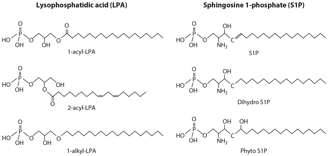
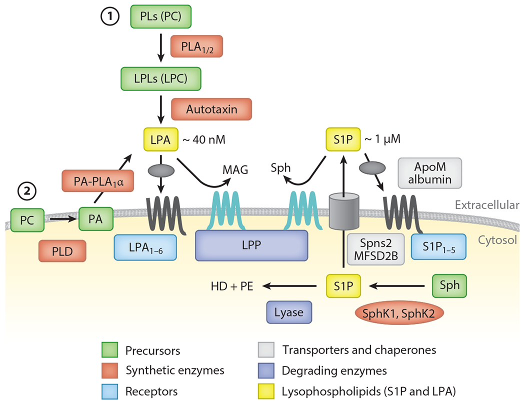
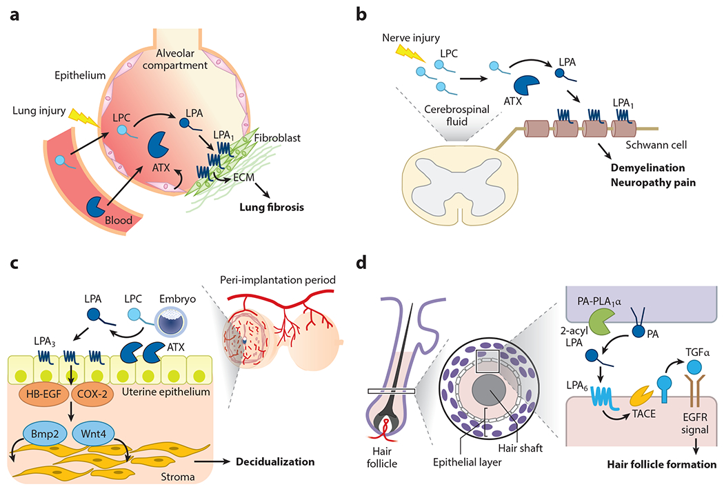
Lysophospholipids, exemplified by lysophosphatidic acid (LPA) and sphingosine 1-phosphate (S1P), are produced by the metabolism and perturbation of biological membranes. Both molecules are established extracellular lipid mediators that signal via specific G protein-coupled receptors in vertebrates. This widespread signaling axis regulates the development, physiological functions, and pathological processes of all organ systems. Indeed, recent research into LPA and S1P has revealed their important roles in cellular stress signaling, inflammation, resolution, and host defense responses. In this review, we focus on how LPA regulates fibrosis, neuropathic pain, abnormal angiogenesis, endometriosis, and disorders of neuroectodermal development such as hydrocephalus and alopecia. In addition, we discuss how S1P controls collective behavior, apoptotic cell clearance, and immunosurveillance of cancers. Advances in lysophospholipid research have led to new therapeutics in autoimmune diseases, with many more in earlier stages of development for a wide variety of diseases, such as fibrotic disorders, vascular diseases, and cancer.

Keywords: cancer; fibrosis; immunology; lysophosphatidic acid; lysophospholipids; sphingosine 1-phosphate; vascular biology.


Conflict of interest statement

DISCLOSURE STATEMENT

T.H. is an inventor named on patents and patent applications related to ApoM-Fc, ApoM+-HDL, and S1PR modulators and has consulted for Pfizer Inc., Sandoz Inc., Novartis Inc., SPARC Inc., Bristol Myers Squibb Inc., and Arena Pharmaceuticals Inc. The other authors are not aware of any affiliations, memberships, funding, or financial holdings that might be perceived as affecting the objectivity of this review.

Figures

Figure 1
Figure 1
Structures of lysophosphatidic acid (LPA) and sphingosine 1-phosphate (S1P). LPA is classified as 1-acyl-LPA, 2-acyl-LPA, or 1-alkyl-LPA according to the differences in the linkage of a fatty acid to the glycerol backbone. S1P is classified as S1P, dihydro S1P,or phyto S1P on the basis of differences in the sphingosine backbone. The fatty acid carbons of S1P are not diverse, whereas the fatty acids of LPA are extremely diverse in terms of their length and degree of unsaturation, which results in different biological activities (via receptor activation).
Figure 2
Figure 2
Production, degradation, transport, and action of lysophosphatidic acid (LPA) and sphingosine 1-phosphate (S1P). Whereas LPA is produced extracellularly, S1P is produced intracellularly. At least two pathways are postulated for LPA. (①) In the autotaxin (ATX) pathway, lysophospholipids (LPLs), mainly lysophosphatidylcholine (LPC), are produced by the action of phospholipase A1 or A2 (PLA1/2), and the resulting LPLs are converted to LPA by ATX. (②) In the non-ATX pathway, phosphatidic acid (PA) is generated on the cell membrane, possibly by phospholipase D (PLD), and the resulting PA is converted to LPA by PA-selective phospholipase A1 (PA-PLA1α). LPA targets the six LPA receptors (LPA1–6). LPA is specifically degraded by lipid phosphate phosphatases (LPPs) to monoacylglycerol (MAG) and phosphate. S1P is produced mainly from sphingosine (Sph) by a phosphorylation reaction. Two sphingosine kinases (SphK1 and SphK2) have been identified. S1P produced intracellularly is transported outside by S1P-specific transporters (Spns2 and MFSD2B). Meanwhile, S1P is degraded by lyase to hexadecenal (HD) and phosphoethanolamine (PE). After transport to the extracellular milieu, S1P binds to ApoM on high-density lipoprotein (HDL) and circulates in the bloodstream. LPPs are also responsible for extracellular degradation of S1P. HDL-bound S1P is brought close to the receptors (S1P1–5), which then activates intracellular signaling pathways.
Figure 3
Figure 3
Pathophysiology of lysophosphatidic acid (LPA). LPA signaling via specific LPA-producing enzymes and LPA receptors is involved in pathophysiological conditions, including (a) lung fibrosis, (b) neuropathy pain, (c) uterine decidualization, and (d) hair follicle formation. (a) Upon lung injury, lysophosphatidylcholine (LPC) and autotaxin (ATX) levels increase and activate the LPA1 receptor on fibroblasts in the alveolar compartment, which leads to the progression of fibrosis by depositing extracellular matrix (ECM) components. (b) Upon nerve injury, newly produced LPC is converted to LPA by ATX, which is always present in cerebrospinal fluid. LPA then acts on LPA1 in myelin, inducing demyelination and the subsequent manifestation of pain. (c) When fertilized eggs interact with uteri (implantation), LPC present in eggs is converted to LPA by ATX, which is expressed abundantly on the surface of uteri (uterine epithelium). Accordingly, LPA3 is activated, as the epithelium also expresses a high level of LPA3, which then activates the decidualization factors such as cyclooxygenase-2 (COX-2), heparin-binding epidermal growth factor (HB-EGF), Wnt4, and Bmp2. Ectopic activation of LPA3 also induces endometriosis (not shown). (d) An LPA-producing enzyme, phosphatidic acid (PA)-selective phospholipase A1 (PA-PLA1α), and an LPA receptor, LPA6, are expressed in specific layers of keratinocytes in hair follicles. Activation of LPA6 induces an ectodomain shedding of transforming growth factor α (TGFα), an epidermal growth factor receptor (EGFR) ligand in the skin, which leads to the formation of hair follicles.
Figure 4
Figure 4
Mass spectrometry imaging of lysophosphatidic acid (LPA) and sphingosine 1-phosphate (S1P). The molecular imaging of LPA and S1P by matrix-assisted laser desorption ionization (MALDI)–mass spectrometry imaging (MSI) is combined with on-tissue derivatization using Phos-tag. LPA and S1P form a complex with Phos-tag, which increases detection sensitivity and selectivity of LPA and S1P in MALDI-MSI analysis. The experimental procedure is as follows: The tissue cryosections are sprayed with Pho-tag. The matrix, organic substances facilitating the ionization, is deposited and then MALDI-MSI analysis is performed. After data processing with spatial information, the distributions of LPA and S1P are visualized as an ion intensity of the Phos-tag complex.

References

    1. Moolenaar WH, van Meeteren LA, Giepmans BN. 2004. The ins and outs of lysophosphatidic acid signaling. Bioessays 26:870–81 - PubMed
    1. Kihara Y, Maceyka M, Spiegel S, Chun J. 2014. Lysophospholipid receptor nomenclature review: IUPHAR Review 8. Br. J. Pharmacol 171:3575–94 - PMC - PubMed
    1. Aikawa S, Hashimoto T, Kano K, Aoki J. 2015. Lysophosphatidic acid as a lipid mediator with multiple biological actions. J. Biochem 157:81–89 - PubMed
    1. Cartier A, Hla T. 2019. Sphingosine 1-phosphate: lipid signaling in pathology and therapy. Science 366:eaar5551. - PMC - PubMed
    1. Engelbrecht E, MacRae CA, Hla T. 2021. Lysolipids in vascular development, biology, and disease. Arterioscler. Thromb. Vasc. Biol 41(2):564–84 - PMC - PubMed

Publication types