. 2022 Jan 11;7(1):e141344.
doi: 10.1172/jci.insight.141344.

p300 or CBP is required for insulin-stimulated glucose uptake in skeletal muscle and adipocytes

Affiliations

p300 or CBP is required for insulin-stimulated glucose uptake in skeletal muscle and adipocytes

Vitor F Martins et al. JCI Insight. .

Abstract

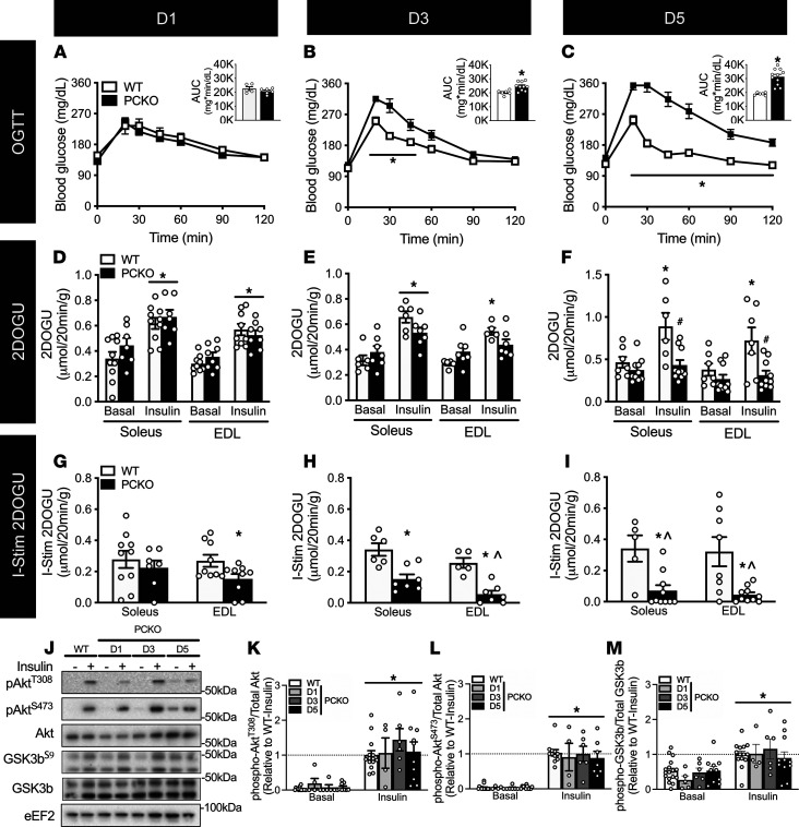
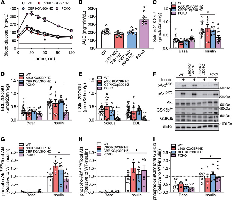
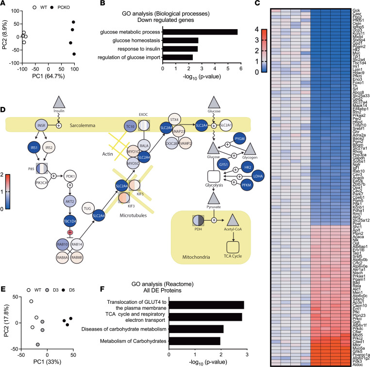
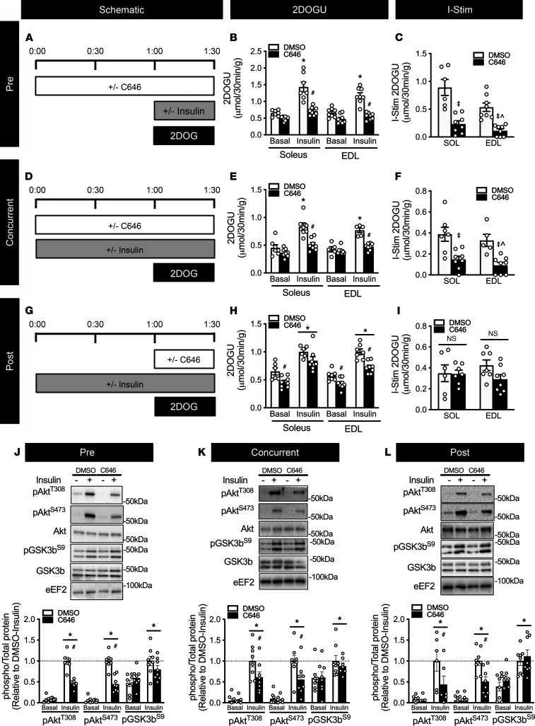
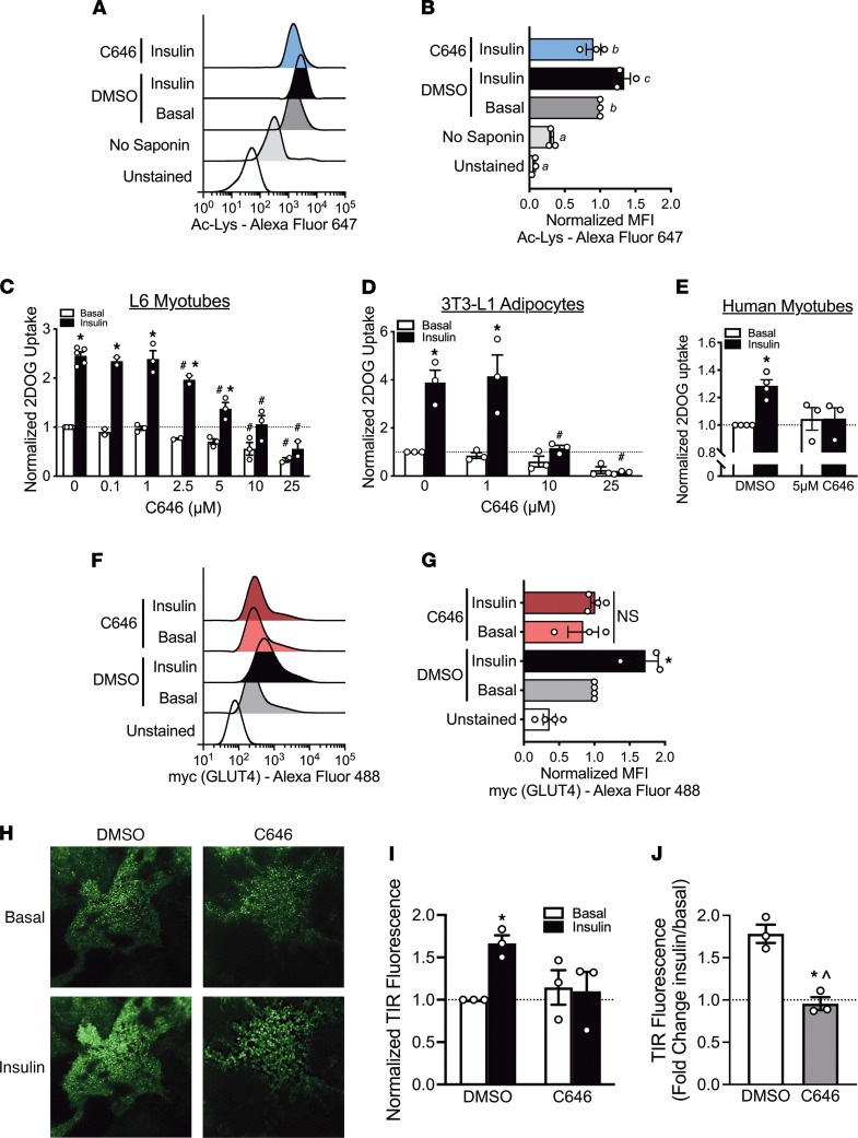
While current thinking posits that insulin signaling to glucose transporter 4 (GLUT4) exocytic translocation and glucose uptake in skeletal muscle and adipocytes is controlled by phosphorylation-based signaling, many proteins in this pathway are acetylated on lysine residues. However, the importance of acetylation and lysine acetyltransferases to insulin-stimulated glucose uptake is incompletely defined. Here, we demonstrate that combined loss of the acetyltransferases E1A binding protein p300 (p300) and cAMP response element binding protein binding protein (CBP) in mouse skeletal muscle caused a complete loss of insulin-stimulated glucose uptake. Similarly, brief (i.e., 1 hour) pharmacological inhibition of p300/CBP acetyltransferase activity recapitulated this phenotype in human and rodent myotubes, 3T3-L1 adipocytes, and mouse muscle. Mechanistically, these effects were due to p300/CBP-mediated regulation of GLUT4 exocytic translocation and occurred downstream of Akt signaling. Taken together, we highlight a fundamental role for acetylation and p300/CBP in the direct regulation of insulin-stimulated glucose transport in skeletal muscle and adipocytes.

Keywords: Endocrinology; Glucose metabolism; Insulin signaling; Muscle Biology; Skeletal muscle.


Conflict of interest statement

Conflict of interest: The authors have declared that no conflict of interest exists.

Figures

Figure 1
Figure 1. p300 and CBP are required for skeletal muscle insulin-stimulated glucose uptake.
Male PCKO mice were assessed at 1 (D1), 3 (D3), or 5 (D5) days after initiating tamoxifen. (AC) Blood glucose concentrations and area under the curve (AUC; inset) for male PCKO mice at D1, D3, and D5 during an oral glucose tolerance test (OGTT; 2 g/kg); D1: WT/PCKO n = 5/9, D3: WT/PCKO n = 6/10, D5: WT/PCKO n = 6/11. *, P < 0.05 2-way ANOVA repeated measures, PCKO versus WT within a time point for OGTT, and *, P < 0.05 t test for AUC. (DF) Basal 2-deoxy-glucose uptake (2DOGU), insulin (0.36 nmol/L) 2DOGU, and (GI) insulin-stimulated 2DOGU (I-Stim.; calculated as insulin 2DOGU – basal 2DOGU) in isolated soleus and extensor digitorum longus (EDL) muscles from male WT and PCKO mice at D1, D3, and D5; D1: WT/PCKO n = 11/10, D3: WT/PCKO n = 6/7, D5: WT/PCKO n = 7/10. *, P < 0.05 2-way ANOVA with Sidak’s multiple comparison versus basal within genotype (DF) and #, P < 0.05 versus WT within basal or insulin (GI). *, P < 0.05 t test for I-Stim. ^, P > 0.05 1-sample t test versus “0”. (J) Representative images for phospho-AktT308 (pAktT308), phospho-AktS473 (pAktS473), total Akt, phospho-GSK3bS9 (pGSK3bS9), and total GSK3b in basal and insulin-stimulated (- and +, respectively) EDL muscles from WT and PCKO mice at D1, D3, and D5. Quantification of (K) pAktT308, (L) pAktS473, and (M) pGSK3bS9 compared with total respective protein abundance in the EDL muscle; WT/D1/D3/D5 n = 15/5/6/11. Values are presented relative to WT-insulin. *, P < 0.05 2-way ANOVA, main effect of insulin. Data reported as mean ± SEM. For Western blots, there were no significant differences between WT mice at the different time points (D1, D3, and D5); therefore WT data were collapsed.
Figure 2
Figure 2. Mice with a single allele of either p300 or CBP have normal glucose tolerance and skeletal muscle insulin action.
Male WT, p300 KO/CBP HZ, CBP KO/p300 HZ, and PCKO mice were assessed at 5 days after initiating tamoxifen. (A) Blood glucose concentrations and (B) AUC for male WT, p300 KO/CBP HZ, CBP KO/p300 HZ, and PCKO mice during an OGTT (2 g/kg); for WT, p300 KO/CBP HZ, CBP KO/p300 HZ, PCKO n = 18/16/9/7. *, P < 0.05 2-way ANOVA with repeated measures, PCKO versus WT within a time point for OGTT (A); *, P < 0.05 1-way ANOVA, versus WT for AUC (B). (C and D) Basal 2DOGU, insulin (0.36 nmol/L) 2DOGU, and (E) insulin-stimulated 2DOGU (calculated as insulin 2DOGU – basal 2DOGU) in isolated soleus and EDL muscles from male WT, p300 KO/CBP HZ, CBP KO/p300 HZ, and PCKO mice; WT, p300 KO/CBP HZ, CBP KO/p300 HZ, PCKO n = 20/8/7/10. *, P < 0.05 2-way ANOVA with Sidak’s multiple comparison versus basal within genotype and #, P < 0.05 versus WT within basal or insulin (C and D). For I-Stim (E), *, P < 0.05 1-way ANOVA with Tukey’s multiple comparison versus WT. ^, P > 0.05 1-sample t test versus “0.” (F) Representative images for pAktT308, pAktS473, total Akt, pGSK3bS9, and total GSK3b in basal and insulin-stimulated (- and +, respectively) EDL muscles from WT, p300 KO/CBP HZ, CBP KO/p300 HZ, and PCKO mice. Quantification of (G) pAktT308, (H) pAktS473, and (I) pGSK3bS9 compared with total respective protein abundance in the EDL muscle; WT, p300 KO/CBP HZ, CBP KO/p300 HZ, PCKO n = 8/5/5/6. Values are presented relative to WT-insulin. *, P < 0.05 2-way ANOVA, main effect of insulin. #, P < 0.05 2-way ANOVA, multiple comparison versus WT-insulin. Data reported as mean ± SEM. For all data, there were no significant differences between WT mice for the respective lines (p300 KO/CBP HZ, CBP KO/p300 HZ, and PCKO); therefore, WT data were collapsed.
Figure 3
Figure 3. Loss of p300/CBP in skeletal muscle leads to the downregulation of insulin signaling and GLUT4 exocytic translocation genes and proteins.
(A) Principal component analysis (PCA) plot for gene expression during microarray in WT and PCKO EDL muscles; WT/PCKO n = 4/4. (B) Gene Ontology (GO) biological processes terms relating to metabolism (FDR < 0.1) for significantly downregulated genes in PCKO versus WT muscles. (C) Expression profile (down [blue] and up [red]) for genes within biological processes GO categories from B. (D) Graphical representation of differentially expressed genes within the insulin signaling, GLUT4 exocytic translocation, and glucose metabolism pathways. (E) PCA plot for protein abundance during tandem mass tag mass spectrometry in WT, D3 PCKO, and D5 PCKO tibialis anterior muscle; WT/D3/D5 n = 3/3/3. (F) GO Reactome terms relating to metabolism (FDR < 0.1) for all differentially expressed proteins in PCKO D5 versus WT muscles.
Figure 4
Figure 4. Acute inhibition of p300/CBP activity in skeletal muscle hinders insulin sensitivity independent of Akt activity.
Schematic for (A) “Pre,” (D) “Concurrent,” and (G) “Post” C646 treatment experiments; 50 μM C646 was used for all experiments. Basal 2DOGU, insulin (0.36 nmol/L) 2DOGU, and insulin-stimulated 2DOGU (calculated as insulin 2DOGU – basal 2DOGU) in isolated soleus and EDL muscles from WT mice in (B and C) “Pre,” (E and F) “Concurrent,” and (H and I) “Post” experiments. (JL) Representative images for pAktT308, pAktS473, total Akt, pGSK3bS9, and total GSK3b in basal and insulin-stimulated (- and +, respectively) EDL muscles from DMSO and C646 treatment groups for (J) “Pre,” (K) “Concurrent,” and (L) “Post” experiments; values are presented relative to DMSO-insulin. *, P < 0.05, 2-way ANOVA with Sidak’s multiple comparison versus basal within treatment. #, P < 0.05 versus DMSO within insulin. For I-Stim, , P < 0.05, 2-way ANOVA, main effect of diet. ^, P > 0.05, 1-sample t test versus “0.” DMSO/C646 n = 8/8. Data reported as mean ± SEM.
Figure 5
Figure 5. p300/CBP activity is required for insulin-stimulated acetylation and GLUT4 trafficking.
(A) Representative experiment and (B) normalized mean fluorescence intensity for total acetylated proteins (Ac-Lys) in L6 myoblasts in unstained, stained without saponin, DMSO, 100 nM insulin, or insulin cotreated with 10 μM C646 conditions, as analyzed by flow cytometry. Different letters signify P < 0.05 1-way ANOVA, Tukey’s multiple-comparison test. All results are representative of 3 independent experiments. Normalized basal and insulin (100 nM) 2DOGU in (C) L6 myotubes, (D) 3T3-L1 adipocytes, and (E) human skeletal muscle myotubes pretreated for 1 hour with DMSO or C646. *, P < 0.05 2-way ANOVA with Sidak’s multiple comparison versus basal within C646 concentration. #, P < 0.05 2-way ANOVA with Sidak’s multiple comparison versus DMSO within insulin treatment. All results are representative of 3 independent experiments. (F) Representative experiment and (G) normalized mean fluorescence intensity for plasma membrane localized GLUT4 in L6-GLUT4-myc myoblasts in unstained, and with or without 25 μM C646 or 100 nM insulin, as analyzed by flow cytometry. *, P < 0.05 2-way ANOVA with Sidak’s multiple comparison versus basal within treatment. Results are representative of 4 independent experiments. (H) Representative total internal reflection fluorescence imaging, using a 60× oil objective (n.a. 1.45), of Myc7-Glut4-eGFP in 3T3-L1 adipocytes 20 minutes after insulin administration and (I and J) its normalized quantitation. (I) *, P < 0.05 2-way ANOVA with Sidak’s multiple comparison versus basal within treatment. (J) *, P < 0.05 t test versus DMSO. ^, P > 0.05 1-sample t test versus “1.” Results are representative of 3 independent experiments with 6–11 cells per experiment. Data reported as mean ± SEM.

Similar articles

Cited by

References

    1. Leto D, Saltiel AR. Regulation of glucose transport by insulin: traffic control of GLUT4. Nat Rev Mol Cell Biol. 2012;13(6):383–396. doi: 10.1038/nrm3351. - DOI - PubMed
    1. Klip A, et al. Thirty sweet years of GLUT4 J Biol Chem. 2019;294(30):11369–11381. - PMC - PubMed
    1. Choudhary C, et al. Lysine acetylation targets protein complexes and co-regulates major cellular functions. Science. 2009;325(5942):834–840. doi: 10.1126/science.1175371. - DOI - PubMed
    1. Kim SC, et al. Substrate and functional diversity of lysine acetylation revealed by a proteomics survey. Mol Cell. 2006;23(4):607–618. doi: 10.1016/j.molcel.2006.06.026. - DOI - PubMed
    1. Lundby A, et al. Proteomic analysis of lysine acetylation sites in rat tissues reveals organ specificity and subcellular patterns. Cell Rep. 2012;2(2):419–431. doi: 10.1016/j.celrep.2012.07.006. - DOI - PMC - PubMed

Publication types