

Association between subtalar articular surface typing and flat foot deformity: which type is more likely to cause flat foot deformity
- PMID: 34814890
- PMCID: PMC8611995
- DOI: 10.1186/s12891-021-04872-8
Association between subtalar articular surface typing and flat foot deformity: which type is more likely to cause flat foot deformity
Abstract
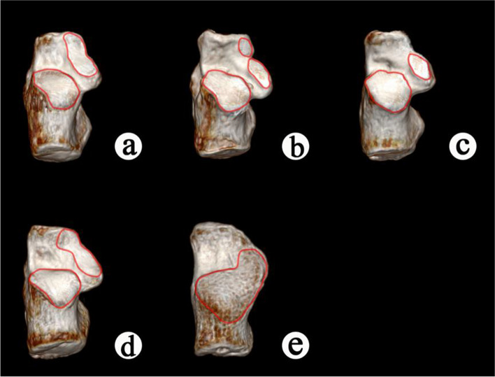
Background: Previous studies have shown a wide range of anatomical classifications of the subtalar joint (STJ) in the population and this is related to the different force line structures of the foot. Different subtalar articular surface morphology may affect the occurrence and development of flat foot deformity, and there are fewer studies in this area. The main objective of our study was to determine the association of different subtalar articular surface with the occurrence and severity of flat foot deformity.
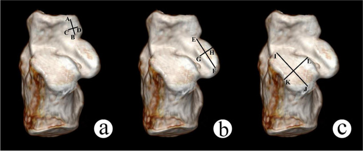
Methods: We analyzed the imaging data of 289 cases of STJ. The articular surface area, Gissane's angle and Bohler's angle of subtalar articular surface of different types were counted. The occurrence and severity of flat foot deformity in different subtalar articular surface were judged by measuring the Meary angle of foot.
Results: We classified 289 cases of subtalar articular surface into five types according to the morphology. According to Meary angle, the flat foot deformity of Type I and Type IV are significantly severer than Type II (P < 0.05). Type II (7.65 ± 1.38 cm2) was significantly smaller than Type I (8.40 ± 1.79 cm2) in the total joint facet area(P < 0.05). Type III (9.15 ± 1.92 cm2) was smaller than Type I (8.40 ± 1.79 cm2), II (7.65 ± 1.38 cm2) and IV (7.81 ± 1.74 cm2) (P < 0.05). Type II (28.81 ± 7.44∘) was significantly smaller than Type I (30.80 ± 4.61 degrees), and IV (32.25 ± 5.02 degrees) in the Bohler's angle (P < 0.05). Type II (128.49 ± 6.74 degrees) was smaller than Type I (131.58 ± 7.32 degrees), and IV (131.94 ± 5.80 degrees) in the Gissane's angle (P < 0.05).
Conclusions: After being compared and analyzed the measurement of morphological parameters, joint facet area and fusion of subtalar articular surface were closely related to the severity of flat foot deformity and Type I and IV were more likely to develop severer flat foot deformity.
Level of evidence: Level III, retrospective comparative study.
Keywords: Flat foot; Meary angle; Subtalar joint.
© 2021. The Author(s).
Conflict of interest statement
The authors declare that they have no competing interests.
Figures
References
-
- Navarro-Flores E, Losa-Iglesias ME, Becerro-de-Bengoa-Vallejo R, Reina-Bueno M, Lopez-Lopez D, Romero-Morales C, et al. Cross-cultural adaptation, translation, and validation of the Spanish foot and ankle outcome score questionnaire. Int Wound J. 2020;17(5):1384–1390. doi: 10.1111/iwj.13400. - DOI - PMC - PubMed
MeSH terms
LinkOut - more resources
Full Text Sources
Research Materials
