. 2021 Nov 30;118(48):e2111946118.
doi: 10.1073/pnas.2111946118.

BAP1 forms a trimer with HMGB1 and HDAC1 that modulates gene × environment interaction with asbestos

Affiliations

BAP1 forms a trimer with HMGB1 and HDAC1 that modulates gene × environment interaction with asbestos

Flavia Novelli et al. Proc Natl Acad Sci U S A. .

Abstract

Carriers of heterozygous germline BAP1 mutations (BAP1+/-) are affected by the "BAP1 cancer syndrome." Although they can develop almost any cancer type, they are unusually susceptible to asbestos carcinogenesis and mesothelioma. Here we investigate why among all carcinogens, BAP1 mutations cooperate with asbestos. Asbestos carcinogenesis and mesothelioma have been linked to a chronic inflammatory process promoted by the extracellular release of the high-mobility group box 1 protein (HMGB1). We report that BAP1+/- cells secrete increased amounts of HMGB1, and that BAP1+/- carriers have detectable serum levels of acetylated HMGB1 that further increase when they develop mesothelioma. We linked these findings to our discovery that BAP1 forms a trimeric protein complex with HMGB1 and with histone deacetylase 1 (HDAC1) that modulates HMGB1 acetylation and its release. Reduced BAP1 levels caused increased ubiquitylation and degradation of HDAC1, leading to increased acetylation of HMGB1 and its active secretion that in turn promoted mesothelial cell transformation.

Keywords: HMGB1; asbestos; gene × environment; germline BAP1 mutations; mesothelioma.


Conflict of interest statement

Competing interest statement: M.C. has a patent issued for BAP1. M.C. and H.Y. have two patents issued for HMGB1. M.C. is a board-certified pathologist who provides consultation for pleural pathology, including medical–legal.

Figures

Fig. 1.
Fig. 1.
BAP1 binds to HMGB1. (A) PLA showing the interaction of BAP1 and HMGB1 (red dots) in the nuclei of BAP1WT and BAP1+/− fibroblasts (nuclei stained blue with DAPI); (Scale bar: 10 μM.) (B) Bar graph: Quantification of PLA red dots per cell showing reduced BAP1–HMGB1 interaction in BAP1+/− fibroblasts compared to BAP1WT. Data shown are mean ± SD (n = 6 cells). P value was calculated using two-tailed unpaired Welch's t test, ***P < 0.001. (C) Schematic representation of full-length BAP1 (1 to 729 aa), the predicted truncations of BAP1 resulting from the germline mutations in the L (1 to 683 aa) and W (1 to 193 aa) families, and the different BAP1 domains: N-terminal ubiquitin carboxy (C)-terminal hydrolase (UCH) domain (aa 1 to 240), a NORS domain (aa 240 to 598), a CTD (aa 598 to 699), and a NLS (aa 699 to 729). Numbers refer to amino acid positions, see also ref. . (D) Co-IP: HEK293 cells were cotransfected with Flag-HMGB1 and the indicated Myc-tagged BAP1 expression vectors; cell extracts were used for Co-IP with anti-Flag resin. Both Myc-tagged BAP1 and its catalytically inactive isoform Myc-BAP1(C91S) interact with Flag-HMGB1, whereas the mutant constructs Myc-BAP1(L) and Myc-BAP1(W) lose the interaction with Flag-HMGB1. The BAP1 domain CTD-NLS had the highest binding affinity to Flag-HMGB1.
Fig. 2.
Fig. 2.
Reduced BAP1 levels increase HMGB1 secretion. (A) Immunofluorescence: HMGB1 localization in BAP1WT and BAP1+/− fibroblasts transfected with H2B-RFP (nuclear marker, red) and immune stained for HMGB1 (green). Merged images show the overlapping yellow signal between HMGB1 and H2B-RFP. The intensity of extranuclear HMGB1 was highlighted by adding a rainbow RGB (red, green, blue) LUT (lookup table) to the merged images. (Scale bar: 10 μM.) (B and C) Bar graphs: Quantification of extranuclear (B) and nuclear (C) HMGB1 measured as fluorescence intensity. Higher levels of extranuclear HMGB1 are seen in BAP1+/− fibroblasts, while higher levels of nuclear HMGB1 are seen in BAP1WTfibroblasts. Data shown are mean ± SD (n = 10 cells per condition). (D) ELISA: Significantly higher HMGB1 levels in conditioned cell culture media of BAP1+/− fibroblasts compared to BAP1WT; experiments were performed in technical duplicates, data shown are mean ± SD of n = 4 biological replicates of BAP1WT and BAP1+/− fibroblasts matched by gender and age. (E) Immunoblot: Amounts of HMGB1 in the total cell lysate and in the conditioned cell culture media of BAP1WT and BAP1+/− fibroblasts; increased HMGB1 levels were detected in the cell culture media of BAP1+/− fibroblasts compared to BAP1WT. (F and G) Total cell lysate and conditioned media were analyzed by immunoblot in HMs (F) and in macrophages (G) transfected with control scramble siRNA, or siBAP1 (siBAP1#1 and siBAP1#5); BAP1 silencing leads to an increased extracellular HMGB1 release in the conditioned media in all cellular models analyzed. (H) Total cell lysate and conditioned media were analyzed by immunoblot in different pairs of BAP1WT and BAP1+/− fibroblasts from L family members; increased levels of actively secreted acetyl-Lys12 HMGB1 and HMGB1 were detected in the conditioned cell culture media of BAP1+/− compared to BAP1WT fibroblasts. (I and J) Immunoblot of serum samples collected from L (I) and W family members (J). (I) Healthy L individuals matched by gender and age: 9_BAP1WT and 13_BAP1+/−. (J) Healthy W individuals matched by gender and age: 26_BAP1WT and 21_BAP1+/−, and one mesothelioma (MM) patient in remission 04_BAP1+/− MM(−) and when relapsed 10 y later, MM(+). Increased levels of acetyl-Lys12 HMGB1 and HMGB1 were present in sera of germline BAP1+/− mutation carriers, and an even greater increase was detected after mesothelioma relapsed. (K) Immunoblot of serum samples collected from healthy Bap1WT and Bap1+/− mice, before and after (2 or 4 mo) asbestos injection, Asb(2m), Asb(4m), and 1 y after injection when the mouse developed MM [Asb(1y), MM]. Increased serum levels of acetyl-Lys12 HMGB1 were detected in the mouse carrying germline Bap1+/− mutations. Further increase was detected after asbestos exposure, and even more after MM developed. Decimals: EG, HMGB1/Ponceau S; HK, acetyl-Lys12 HMGB1/Ponceau S and HMGB1/Ponceau; P values were calculated using two-tailed unpaired Welch's t test, *P < 0.05, ***P < 0.001.
Fig. 3.
Fig. 3.
BAP1 decreases HMGB1 secretion and malignant transformation. (A–C) In vitro transformation measured as tridimensional foci formation. Primary HM cells were silenced with scramble siRNA or a pool of siBAP1, and treated with 100 ng/mL Box A (A), or 10 mM EP (B), or 100 μM aspirin (C), and then exposed to crocidolite asbestos (5 μg/cm2) in the presence of TNFα. Inhibition of HMGB1 secretion with Box A, EP, or aspirin, decreased foci formation in BAP1-silenced HMs. Data shown are mean ± SE of n = 9 technical replicates from n = 3 independent experiments (A and B), and of n = 6 technical replicates from n = 2 independent experiments (C). (D) Aspirin reduces the secretion of acetyl-Lys12 HMGB1 and HMGB1 in the conditioned cell culture media of HM cells transfected with a pool of siBAP1. Decimals: acetyl-Lys12 HMGB1/Ponceau S, HMGB1/Ponceau S. (E and F) Mesothelioma incidence (E) and mesothelioma-specific survival (F) in aspirin-treated Bap1+/− mice after exposure to crocidolite. For mesothelioma incidence, P value was calculated using Fisher's exact test (P = 0.0130); survival curves were compared using Log-rank test (P = 0.0452). P values were calculated using two-tailed unpaired Welch's t test, **P < 0.01, *P < 0.05. We had to exclude 12 and 10 mice from each group, respectively, from analysis because the cause of death was due to factors unrelated to asbestos exposure.
Fig. 4.
Fig. 4.
BAP1 prevents HDAC1 degradation. (A) Fluorimetric assay. Reduced HDAC activity in fibroblasts carrying germline BAP1+/− mutations. Data of in vitro HDAC activity assay on nuclear extracts are displayed as mean ± SE of n = 7 biological replicates of BAP1WT and BAP1+/− fibroblasts matched by gender and age (n = 4 from W and n = 3 from L family members). Trichostatin A (TSA), a HDAC inhibitor, was utilized to check the specificity of the HDAC activity assay. P value was calculated using two-tailed unpaired Welch's t test, *P < 0.05. (B) Immunoblot of HDAC1 and HDAC4 protein levels in total cell lysates of BAP1WT and BAP1+/− fibroblasts showing reduced HDAC1 in BAP1+/− compared to BAP1WT fibroblasts. Decimals: HDAC1/α-tubulin. (C) Immunoblot: Reintroduction of HDAC1 decreases the secretion of HMGB1 and acetyl-Lys12 HMGB1 in the conditioned cell culture media of BAP1+/− fibroblasts compared to control cells. Decimals: Acetyl-Lys12 HMGB1/Ponceau S and HMGB1/Ponceau S. (D) In vitro ubiquitylation assay. HEK293 cells were cotransfected with Flag-tagged HDAC1 and Myc-tagged wild-type BAP1, the catalytically inactive BAP1(C91S), BAP1(L) and BAP1(W), pCMV6-AN-Myc was used as a negative control. Wild-type BAP1 interacts and deubiquitylates HDAC1 compared to BAP1(C91S); truncated BAP1(L) and BAP1(W) are also able to interact and deubiquitylate HDAC1 in vitro. Decimals: Ubiquitin/HDAC1. (E) Mapping of the BAP1 region interacting with HDAC1. HEK293 cells were cotransfected with Flag-HDAC1 and the indicated Myc-tagged BAP1 expression vectors (see also Fig. 1C); cell extracts were used for Co-IP with anti-Flag resin. The UCH domain of BAP1 had the highest binding affinity to HDAC1, while the NORS region alone showed no interaction, and the CTD-NLS region only slightly contributed to the binding.
Fig. 5.
Fig. 5.
Characterization of HMGB1 binding to BAP1 and HDAC1. (A) Co-IP of endogenous BAP1 and HDAC1 with HMGB1 (used as bait) in fibroblast cell cultures from BAP1WT individuals or carriers of heterozygous BAP1+/− mutations. Higher amounts of the coprecipitated BAP1–HDAC1 proteins are found in BAP1WT cells. (B) Schematic representation of HMGB1 domains. HMGB1 (1 to 215 amino acids) consists of an N-terminal A box domain (aa 1 to 79), a B box domain (aa 89 to 162), and a C-terminal acidic tail (aa 186 to 215). Numbers refer to amino acid (aa) positions. (C) Mapping of the HMGB1 region interacting with BAP1. HEK293 cells were cotransfected with Myc-tagged BAP1 and the Flag-tagged HMGB1 fragments displayed in B. Cell extracts were used for Co-IP with anti-Flag resin. The HMGB1-A/B box domain had the strongest binding affinity to Myc-tagged BAP1; A box and B box domains alone showed weaker interaction, and the B box-Ctail region slightly contributed to the binding. (D) Mapping of the HMGB1 region interacting with HDAC1. HEK293 cells were cotransfected with the indicated Flag-tagged HMGB1 fragments (B). Cell extracts were used for Co-IP with an anti-Flag resin. The HMGB1-A/B box domain showed the strongest binding affinity to endogenous HDAC1.
Fig. 6.
Fig. 6.
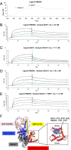
BAP1, HDAC1, and HMGB1 form a trimeric protein complex. (A) SPR sensorgram showing analyte binding of HDAC1 and BAP1 (167 nM), sequentially passed over immobilized HMGB1 without intermediate chip surface regeneration. (B–D) Sensorgrams and fitted curves, applying a Langmuir 1:1 binding model. Analytes were bound in serial dilutions (red: 167 nM, blue: 84 nM, green: 42 nM, turquoise: 21 nM) to the ligand covalently immobilized on the surface of a Biacore CM5 sensor chip. Binding of BAP1 to HMGB1 (B); binding of HDAC1 to BAP1 (C); binding of HDAC1 to HMGB1 (D). (E) HDAC1 and BAP1 were coincubated in serial dilutions (red: 167 nM, blue: 84 nM, green: 42 nM, turquoise: 21 nM) for 10 min in HBS-EP buffer. The preformed complexes were subsequently passed as analyte over the immobilized HMGB1 ligand. Kinetic constants and affinity were determined from the concentration sensorgrams by Langmuir 1:1 curve fitting. The equilibrium dissociation constants (KD; in nanomoles) are shown. (F) Structural model of the BAP1–HMGB1–HDAC1 trimeric protein complex. BAP1: UCH, blue; NORS, pink; CTD, yellow; HMGB1 (A box–B box), red; HDAC1, gray. The binding interface between BAP1 and HMGB1 is zoomed in. The hydrophobic patch between the B box of HMGB1 and BAP1 is highlighted: light blue circle.
Fig. 7.
Fig. 7.
BAP1, HDAC1, and HMGB1 trimer regulates chronic inflammation and cell transformation. Schematic representation showing how BAP1 prevents active HMGB1 release through the stabilization of HDAC1. In BAP1WT individuals, nuclear BAP1 forms a trimer with HMGB1 and HDAC1. BAP1 deubiquitylates and stabilizes HDAC1, which can deacetylate HMGB1, thus preventing its active secretion. In BAP1+/− carriers, reduced BAP1 levels result in the degradation of HDAC1 and destabilization of its binding to HMGB1, leading to increased acetylation of HMGB1. Acetylated HMGB1 is then secreted into the extracellular space and promotes chronic inflammation and cell transformation, contributing to the development of mesothelioma. Aspirin by blocking HMGB1 secretion inhibits mesothelial cell transformation and tumorigenesis. Image was made with BioRender.com.

Similar articles

Cited by

References

    1. Carbone M., et al. , Tumour predisposition and cancer syndromes as models to study gene-environment interactions. Nat. Rev. Cancer 20, 533–549 10.1038/s41568-020-0265-y. (2020). - DOI - PMC - PubMed
    1. Carbone M., et al. , Consensus report of the 8 and 9th Weinman Symposia on Gene x Environment Interaction in carcinogenesis: Novel opportunities for precision medicine. Cell Death Differ. 25, 1885–1904 (2018). - PMC - PubMed
    1. Carbone M., et al. , Mesothelioma: Scientific clues for prevention, diagnosis, and therapy. CA Cancer J. Clin. 69, 402–429 (2019). - PMC - PubMed
    1. Sluis-Cremer G. K., Liddell F. D., Logan W. P., Bezuidenhout B. N., The mortality of amphibole miners in South Africa, 1946-80. Br. J. Ind. Med. 49, 566–575 (1992). - PMC - PubMed
    1. Bianchi M. E., et al. , High-mobility group box 1 protein orchestrates responses to tissue damage via inflammation, innate and adaptive immunity, and tissue repair. Immunol. Rev. 280, 74–82 (2017). - PubMed

Publication types

MeSH terms