. 2021 Nov 23;12(1):6811.
doi: 10.1038/s41467-021-27016-x.

Reversible and spatiotemporal control of colloidal structure formation

Affiliations

Reversible and spatiotemporal control of colloidal structure formation

H Dehne et al. Nat Commun. .

Abstract

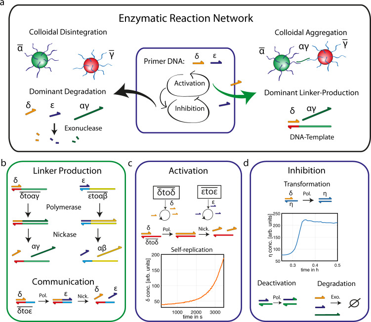
Tuning colloidal structure formation is a powerful approach to building functional materials, as a wide range of optical and viscoelastic properties can be accessed by the choice of individual building blocks and their interactions. Precise control is achieved by DNA specificity, depletion forces, or geometric constraints and results in a variety of complex structures. Due to the lack of control and reversibility of the interactions, an autonomous oscillating system on a mesoscale without external driving was not feasible until now. Here, we show that tunable DNA reaction circuits controlling linker strand concentrations can drive the dynamic and fully reversible assembly of DNA-functionalized micron-sized particles. The versatility of this approach is demonstrated by programming colloidal interactions in sequential and spatial order to obtain an oscillatory structure formation process on a mesoscopic scale. The experimental results represent an approach for the development of active materials by using DNA reaction networks to scale up the dynamic control of colloidal self-organization.


Conflict of interest statement

The authors declare no competing interests.

Figures

Fig. 1
Fig. 1. DNA reaction networks control colloidal aggregation.
a Scheme of an enzymatic reaction network that can inhibit and activate the amplification of the primer DNA strands ε and δ. In the presence of the primer strands, an additional enzymatic reaction is activated and produces a DNA linker strand, which induces the selective structure formation of DNA-coated colloids. b The production of two different types of colloidal linker strands αγ and αβ (24 nT) can be activated by the primer strands ε and δ (10 nT) and results in the specific aggregation of DNA coated colloids, which are fluorescently labeled. In the communication setup, each of the primers is used as a catalytic input to produce the other primer as an output. c The autocatalytic DNA amplification is catalyzed by repetitive DNA templates using a polymerase (Pol.) and a nickase (Nick.) and is monitored using fluorescence spectroscopy (here, δtoδ¯= 120 nM). d The self-replication of the primer strands (ε and δ) can be controlled by three inhibiting reactions. A palindromic DNA sequence η is used to realize the autocatalytic transformation of δ into η (experiment: η = 50 nM and δ = 1 nM). The deactivation of the primer activity is achieved by the addition of a short polynucleotide sequence, and an exonuclease (Exo.) is used to degrade the produced DNA strands.
Fig. 2
Fig. 2. Enzymatic reactions control the speed and composition of colloidal aggregation.
a The aggregation of α¯ and β¯ particles (green) is induced by enzymatic reactions, and the aggregation speed can be tuned by using different template concentrations (δtoδ¯). b The autocatalytic production of ε is used to control the structure formation of α¯ and γ¯ particles and results in clusters of red and green particles. c The two self-replicating reactions can be coupled by using the “communication-reaction” of δtoε¯. The amplification of δ is induced at first, and the time-delay of the subsequent reaction is determined by the concentration of δtoε¯. The delayed production of αγ linker results in a controllable initiation of α¯ and γ¯ aggregation. The programming of the enzymatic setup allows tuning of the colloidal composition, as shown by the green backbone (α¯ and β¯) and the surrounding of red particles (γ¯, scale bar = 10 µm). The time traces of the two reactions are simulated to demonstrate the induced time-delay. The amplification of δ is kept constant, while the concentrations of δtoε¯ are varied to control the initiation of the ε amplification. The time of aggregation was determined at ε = 1 arb. units.
Fig. 3
Fig. 3. Traveling aggregation fronts and communication.
a The local linker production of functionalized particles (Pδtoαβ¯) generates concentration profiles and results in local aggregation (red area) within a microfluidic chamber (~1 cm channel). b Traveling fronts of colloidal aggregation are induced by the local activation and subsequent propagation of the primer strands. The speed of the propagation is tuned by varying the reaction speed by the global concentration of δtoδ¯ strands. The images of the colloidal aggregates along the channel are depicted at t = 90 min. c Signal propagation along the microfluidic channel is realized by the local activation at one microfluidic chamber and the propagation of δ along the channel. The local colloidal structure formation is induced by the activation of the linker production once the signal reaches the functionalized particles (Pδtoαβ¯). d Both self-replicating systems are used to enable the communication between the two microfluidic chambers. The amplification of ε is activated locally, leading to the signal propagation, which is transformed to δ at the end of the channel due to the functionalized particles Pεtoδ¯. The transformation induces a backward signal propagation which is finally coupled to the colloidal aggregation (δtoαβ¯).
Fig. 4
Fig. 4. Oscillating structure formation.
a Scheme of the oscillating reaction network (blue dotted box) and the coupled linker reaction (red dotted boxes). The oscillation of the DNA amplification (blue box) can be controlled in terms of frequency and can activate the linker production during the DNA concentration peaks (red box). Here, one cycle of the oscillation is shown with (green) and without (orange) additional linker production. b Oscillating linker concentrations in dependency of the template concentration and linker production. The green area depicts the phase space, which enables the synchronization of the DNA concentration profiles and the colloidal aggregation. At different points in time, colloids were added (red circles) and the microscopy snapshots demonstrate the corresponding aggregation (i, iii) and disintegration (ii), scale bar = 15 µm. c Colloidal cluster size monitored over time for different template concentrations (see “Methods”, microscopy experiments). The monodisperse state is depicted in blue, while the colloidal clusters are highlighted in red. Bottom: Microscopy snapshots of the colloidal structure formation during 10 h of oscillating linker production (Supplementary Video, δtoδ¯ = 400 nM, with peaks of aggregations at 51, 149, 243 and 557 min, scalebar = 15 µm).

References

    1. Glotzer SC, Solomon MJ, Kotov NA. Self‐assembly: from nanoscale to microscale colloids. AIChE J. 2004;50:2978–2985. doi: 10.1002/aic.10413. - DOI
    1. Khlebtsov NG, Dykman LA, Krasnov YM, Mel’nikov AG. Light absorption by the clusters of colloidal gold and silver particles formed during slow and fast aggregation. Colloid J. 2000;62:765–779. doi: 10.1023/A:1026643111821. - DOI
    1. Cigler, P., Lytton-Jean, A. K., Anderson, D. G., Finn, M. G. & Park, S. Y. DNA-controlled assembly of a NaTl lattice structure from gold nanoparticles and protein nanoparticles. Nat. Mater.10.1038/nmat2877 (2010). - PMC - PubMed
    1. Fan, J. A. et al. Self-assembled plasmonic nanoparticle clusters. Science10.1126/science.1187949 (2010). - PubMed
    1. Simmel FC, Schulman R. Self-organizing materials built with DNA. MRS Bull. 2017;42:913–919. doi: 10.1557/mrs.2017.271. - DOI

Publication types

MeSH terms