. 2021 Nov 23;11(1):22805.
doi: 10.1038/s41598-021-02030-7.

Assessment of histological characteristics, imaging markers, and rt-PA susceptibility of ex vivo venous thrombi

Affiliations

Assessment of histological characteristics, imaging markers, and rt-PA susceptibility of ex vivo venous thrombi

Samuel A Hendley et al. Sci Rep. .

Abstract

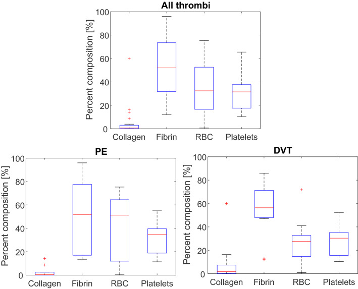
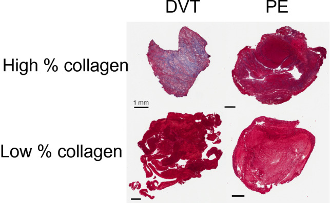
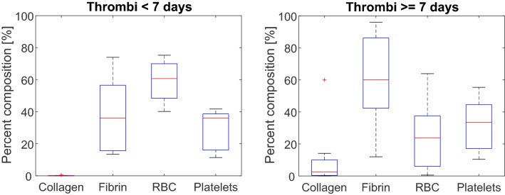
Venous thromboembolism is a significant source of morbidity and mortality worldwide. Catheter-directed thrombolytics is the primary treatment used to relieve critical obstructions, though its efficacy varies based on the thrombus composition. Non-responsive portions of the specimen often remain in situ, which prohibits mechanistic investigation of lytic resistance or the development of diagnostic indicators for treatment outcomes. In this study, thrombus samples extracted from venous thromboembolism patients were analyzed ex vivo to determine their histological properties, susceptibility to lytic therapy, and imaging characteristics. A wide range of thrombus morphologies were observed, with a dependence on age and etymology of the specimen. Fibrinolytic inhibitors including PAI-1, alpha 2-antiplasmin, and TAFI were present in samples, which may contribute to the response venous thrombi to catheter-directed thrombolytics. Finally, a weak but significant correlation was observed between the response of the sample to lytic drug and its magnetic microstructure assessed with a quantitative MRI sequence. These findings highlight the myriad of changes in venous thrombi that may promote lytic resistance, and imaging metrics that correlate with treatment outcomes.


Conflict of interest statement

K.B.B. obtains funding from the National Institutes of Health (Grant Number R01 HL133334). O. A. has acted as a consultant for Inari Medical, Boston Scientific, and received research grants from Inari Medical, Canon Medical, and Philips. He acted as a speaker and received compensation for Argon Medical, Canon Medical, Penumbra, Philips, and Johnson and Johnson. G.D.W. received honoraria and serves on the advisory board for Diagnostica Stago. S.A.H., A.B., A.D., E.S., D.M., R.O.A., Y.Z., S.S., Z.F.L., J.D.P. declare no potential conflict of interest.

Figures

Figure 1
Figure 1
The fractional area of collagen, fibrin, platelets, and erythrocytes (RBC) present in (top) all analyzed VTE specimen (26 samples), (bottom left) pulmonary emboli (14 samples), and (bottom right) deep vein thrombi (12 samples). Components were assessed using histochemical staining. Colorimetric analysis was conducted in ImageScope (Leica, Biosystems, Germany) to identify pixels associated with each components. Horizontal red lines indicate median values. The top and bottom portions of the blue box represent the 25th and 75th percentiles, respectively, and whiskers extent to the data points not considered outliers. Red crosses indicate outliers, which correspond to datapoints greater than q75+1.5×q75-q25 or less than q25-1.5×q75-q25, where q25 corresponds to the 25 th percentile of the sample data, and q75 corresponds 75th percentile of the sample data.
Figure 2
Figure 2
Observed patient-to-patient variability in collagen observed in ex vivo thrombus samples. Deep vein thrombi (DVT) are shown in the left column, pulmonary emboli (PE) are shown in the right column. Collagen appears blue in the Masson’s trichrome stain and compromises 60% (top left) and less than 1% (bottom left) for DVT, and 14% (top right) and less than 1% (bottom right) for PE by area. Colorimetric analysis was conducted in ImageScope (Leica Biosystems, Germany) to identify pixels associated with collagen. Color thresholds were evaluated and accepted by a board-approved pathologist. The scale bars indicate 1 mm.
Figure 3
Figure 3
Trends in thrombus composition for fibrin, collagen, red blood cells (RBCs), and platelets for all samples (N = 26 total). The Pearson’s correlation coefficient (R) and p-values are reported for each respective pair.
Figure 4
Figure 4
Histological analysis for acute (less than 7 days old, N = 12) and chronic (greater than or equal to 7 days old, N = 14) thrombi. None of the 26 samples were excluded from analysis. Red crosses indicate outliers and horizontal red lines indicate median values. The top and bottom portions of the blue box represent the 25th and 75th percentiles, respectively, and whiskers extent to the data points not considered outliers. Outliers correspond to datapoints greater than q75+1.5×q75-q25 or less than q25-1.5×q75-q25, where q25 corresponds to the 25th percentile of the sample data, and q75 corresponds 75th percentile of the sample data .
Figure 5
Figure 5
Representative multiplexed immunofluorescent images collected from VTE sample. (A) DAPI (nucleated cells) in blue, (B) erythrocytes (pink), (C) CD61 (platelets, red), (D) collagen, types i, ii, and iii (orange), (E) fibrin (green), (F) PAI-1 (cyan), and (G) VEGR-1 (yellow). The scale bar in the lower left corner corresponds to a 20 µm distance (40 × magnification). Antibody information can be found in Supplemental Table 2. A total of eight different antibodies were used for analysis, with five shown here. Autofluorescence was used to visualize erythrocytes. No cross reactivity was observed for antibodies as assessed with human tonsil control samples.
Figure 6
Figure 6
(Top) Representative multiplexed thrombus image indicating nucleated cells assessed with DAPI (blue), PAI-1 (yellow) and CD31 (red). Orange arrows indicate representative locations for cells associated with revascularization (co-expression of DAPI and CD31). Magenta arrows indicate representative areas for co-expression of PAI-1 and CD31. The white bar is a 10 µm distance (40 × magnification). Multiplex IHC staining was performed on 5 micron FFPE tissue sections using the Opal 7-Color Manual IHC Kit (NEL861001KT; Akoya Biosciences) following the manufacturer instructions. All slides were counterstained with Spectral DAPI (1:10, Akoya Biosciences) nuclear stain and mounted with Slowfade Diamond Antifade mountant (Thermo Fisher Scientific). The slides were then scanned using the Vectra Polaris whole slides scanner (Akoya Biosciences), the regions of interest were selected using the Phenochart software (Akoya Biosciences) and analyzed using Inform software (Akoya Biosciences). Antibody information can be found in Supplemental Table 2. The individual channels for each components are shown in the lower three panels.
Figure 7
Figure 7
Comparison of sample thrombolytic susceptibility (mass loss) and magnetic susceptibility (QSM analysis). Error bars represent the range of positive QSM values observed within the sample. Separate segments of each specimen were subjected to the clot mass loss assay and imaging analysis. One sample was highly heterogenous (red cross), and gross observation indicated strong variation between the portion of that specimen that was subjected to rt-PA and the portion subjected to QSM analysis. The dashed yellow line is a least-squares best fit to the data. A significant correlation was observed between the thrombolytic and magnetic susceptibility of the sample (Spearman’s rho = 0.90, p < 0.01, N = 8 samples) The heterogenous sample (red cross) was excluded from analysis.

References

    1. Aparicio HJ, et al. Heart disease and stroke statistics-2021 update a report from the American Heart Association. Circulation. 2021 doi: 10.1161/CIR.0000000000000950. - DOI - PubMed
    1. Raskob GE, et al. Thrombosis: A major contributor to global disease burden. Arterioscler. Thromb. Vasc. Biol. 2014;34:2363–2371. - PubMed
    1. Grosse SD. Incidence-based cost estimates require population-based incidence data. A critique of Mahan et al. Thromb. Haemost. 2012;107:192–193. - PubMed
    1. Go AS, et al. Heart disease and stroke statistics–2013 update: A report from the American Heart Association. Circulation. 2013;127:e6–e245. - PMC - PubMed
    1. Fleck D, et al. Catheter-directed thrombolysis of deep vein thrombosis: Literature review and practice considerations. Cardiovasc. Diagn. Ther. 2017;7:S228–S237. - PMC - PubMed

Publication types

MeSH terms