. 2021 Nov 23;11(1):22731.
doi: 10.1038/s41598-021-02210-5.

Feasibility study of portable multi-energy computed tomography with photon-counting detector for preclinical and clinical applications

Affiliations

Feasibility study of portable multi-energy computed tomography with photon-counting detector for preclinical and clinical applications

Chang-Lae Lee et al. Sci Rep. .

Abstract

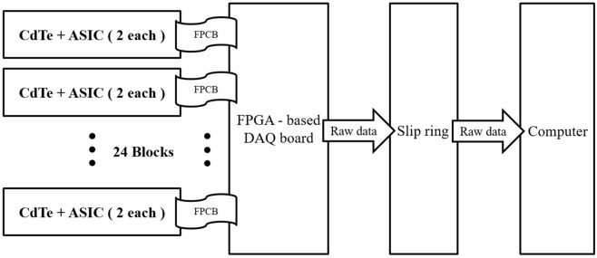
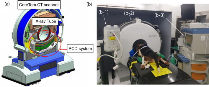
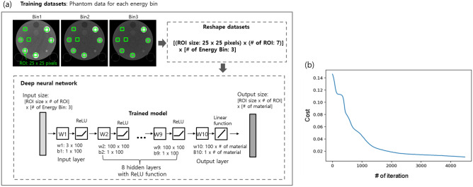
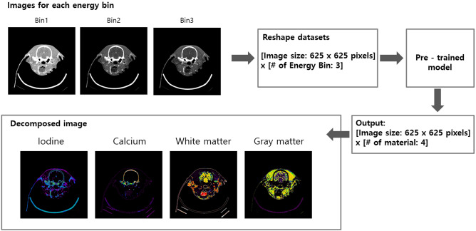
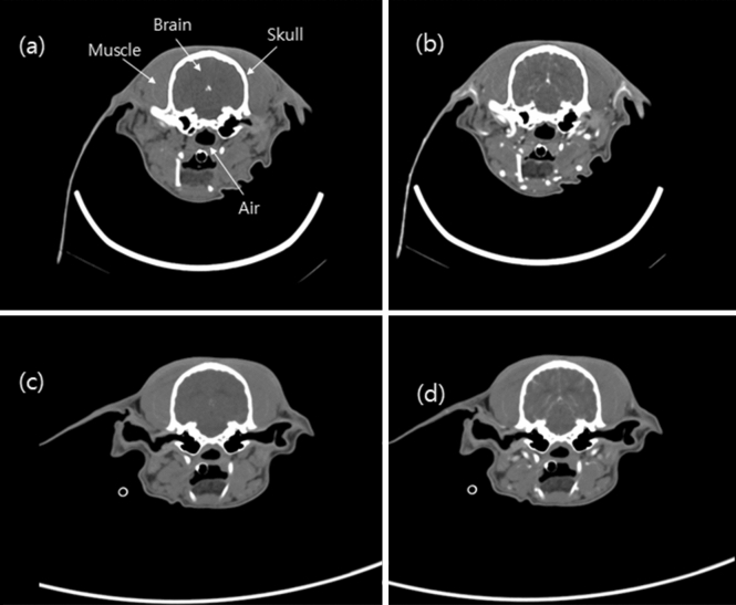
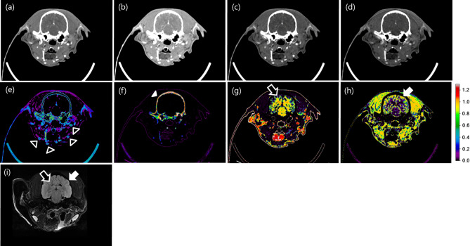
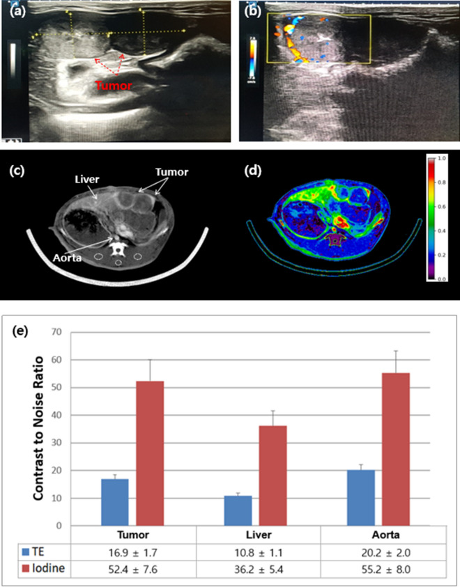
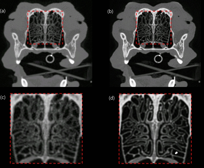
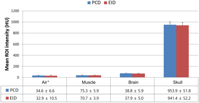
In this study, preclinical experiments were performed with an in-house developed prototypal photon-counting detector computed tomography (PCD CT) system. The performance of the system was compared with the conventional energy-integrating detector (EID)-based CT, concerning the basic image quality biomarkers and the respective capacities for material separation. The pre- and the post-contrast axial images of a canine brain captured by the PCD CT and EID CT systems were found to be visually similar. Multi-energy images were acquired using the PCD CT system, and machine learning-based material decomposition was performed to segment the white and gray matters for the first time in soft tissue segmentation. Furthermore, to accommodate clinical applications that require high resolution acquisitions, a small, native, high-resolution (HR) detector was implemented on the PCD CT system, and its performance was evaluated based on animal experiments. The HR acquisition mode improved the spatial resolution and delineation of the fine structures in the canine's nasal turbinates compared to the standard mode. Clinical applications that rely on high-spatial resolution expectedly will also benefit from this resolution-enhancing function. The results demonstrate the potential impact on the brain tissue segmentation, improved detection of the liver tumors, and capacity to reconstruct high-resolution images both preclinically and clinically.


Conflict of interest statement

The authors declare no competing interests. CL.L., K.H., N.K., K.H., S.L., J.P., KY.L., Y.C., M.C. are employees of Samsung Electronics.

Figures

Figure 1
Figure 1
Photon-counting detector system consisting of CdTe, application-specific integrated circuit (ASIC), and various other components.
Figure 2
Figure 2
(a) Illustration of the photon-counting detector computer tomography (PCD CT) system placed inside the CereTom Scanner. (b) Beagle dog positioned for scanning. (b-1) CT injection system for injecting contrast medium. (b-2) PCD CT system. (b-3) Monitoring system for temperature, blood pressure, peripheral oxygen saturation, and end-tidal carbon dioxide.
Figure 3
Figure 3
(a) Illustration of the structure for networks. The model consists of eight hidden layers with ReLU function. Images are shown as examples. (b) Cost function.
Figure 4
Figure 4
An example of application of the network for material decomposition.
Figure 5
Figure 5
(a) Pre- and (b) post-contrast axial image of the canine’s brain in the PCD system (30–140 keV, 10 mAs, CTDIvol: 41.7 mGy, slice thickness: 2.5 mm, display window/level: W/L = 450/100 HU). (c) Pre- and (d) post-contrast axial image of the head in the EID system (140 kVp, 100 mAs, CTDIvol: 41.7 mGy, slice thickness: 2.0 mm, display window: W/L = 450/100 HU).
Figure 6
Figure 6
Contrast-enhanced axial images of the canine brain acquired at (a) energy levels in the range of 30 to 140 keV in the PCD CT system and (b) multi-energy bin 1: 30–50 keV, (c) bin 2: 50–65 keV, and (d) bin 3: 65–140 keV (CT dose index (CTDIvol): 41.7 mGy, display window: W/L = 450/100 HU, slice thickness: 2.5 mm). An image of iodine (e), calcium (f), white matter (g), and gray matter (h) separated with the use of images of the three energy levels (bins 1, 2, and 3). Axial brain MRI (i) of FLAIR sequence. Black arrowheads indicate arteries, the white arrowhead indicates the skull, the black arrow indicates white matter, and the white arrow indicates gray matter.
Figure 7
Figure 7
Rabbit with VX2 tumors located in the anterior part of the liver presented as (a) two-dimensional and (b) three-dimensional flow images produced via Doppler ultrasonography. (c) Contrast-enhanced axial image acquired at the total energy range and (d) iodine map image of the rabbit liver in the PCD CT system. The dotted circles are back muscles. 30–100 keV, 10 mAs, slice thickness = 2.5 mm, WW/WL [450/100]. (e) Contrast-to-noise ratio (mean ± SD) of the PCD CT system for the tumor, liver, and aorta in the total energy range (TE, 30–140 keV) of a rabbit acquired at an energy level in the range of 30–140 keV and the iodine map.
Figure 8
Figure 8
Axial images of the nasal turbinates scanned with the use of the (a) standard mode and (b) high-resolution (HR) mode of the PCD CT system. The average thickness of nasal turbinates (white arrow) is 0.5 mm. (c, d) The zoomed-in ROIs show better delineation of sub-millimeter nasal turbinates using (c) the HR modecompared with (d) the standard mode.
Figure 9
Figure 9
Mean HU values of air, muscle, brain, and skull image acquired from PCD and EID in Fig. 5. *The mean HU unit values for air were added to the value of 1000 HU for easier visualization.

Similar articles

Cited by

References

    1. Shikhaliev P, Xu T, Molloi S. Photon counting CT: Concept and initial results. Med. Phys. 2005;32:427–436. doi: 10.1118/1.1854779. - DOI - PubMed
    1. Gutjahr R, et al. Human imaging with photon-counting-based CT at clinical dose levels: Contrast-to-noise ratio and cadaver studies. Invest. Radiol. 2016;51:421. doi: 10.1097/RLI.0000000000000251. - DOI - PMC - PubMed
    1. Byl A, et al. Photon-counting normalized metal artifact reduction (NMAR) in diagnostic CT. Med. Phys. 2021;48:3572–3582. doi: 10.1002/mp.14931. - DOI - PubMed
    1. Silva J, et al. Resolution characterization of a silicon-based, photon-counting computed tomography prototype capable of patient scanning. J. Med. Imaging (Bellingham) 2019;6:043502. - PMC - PubMed
    1. Ji X, et al. Development of an integrated C-arm interventional imaging system with a strip photon counting detector and a flat panel detector. IEEE Trans. Med. Imaging. 2021 doi: 10.1109/TMI.2021.3095419. - DOI - PubMed

Publication types