. 2021 Nov 23;11(1):22792.
doi: 10.1038/s41598-021-02254-7.

The von Willebrand factor stamps plasmatic extracellular vesicles from glioblastoma patients

Affiliations

The von Willebrand factor stamps plasmatic extracellular vesicles from glioblastoma patients

Quentin Sabbagh et al. Sci Rep. .

Abstract

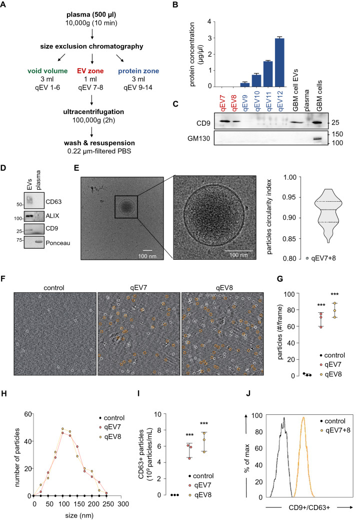
Glioblastoma is a devastating tumor of the central nervous system characterized by a poor survival and an extremely dark prognosis, making its diagnosis, treatment and monitoring highly challenging. Numerous studies have highlighted extracellular vesicles (EVs) as key players of tumor growth, invasiveness and resistance, as they carry and disseminate oncogenic material in the local tumor microenvironment and at distance. However, whether their quality and quantity reflect individual health status and changes in homeostasis is still not fully elucidated. Here, we separated EVs from plasma collected at different time points alongside with the clinical management of GBM patients. Our findings confirm that plasmatic EVs could be separated and characterized with standardized protocols, thereby ensuring the reliability of measuring vesiclemia, i.e. extracellular vesicle concentration in plasma. This unveils that vesiclemia is a dynamic parameter, which could be reflecting tumor burden and/or response to treatments. Further label-free liquid chromatography tandem mass spectrometry unmasks the von Willebrand Factor (VWF) as a selective protein hallmark for GBM-patient EVs. Our data thus support the notion that EVs from GBM patients showed differential protein cargos that can be further surveyed in circulating EVs, together with vesiclemia.


Conflict of interest statement

The authors declare no competing interests.

Figures

Figure 1
Figure 1
Separation of extracellular vesicles from human peripheral blood. (A) Diagram representing the separation of extracellular vesicles (EVs) from peripheral blood. (B) Total protein concentrations were measured in each size-exclusion chromatography (SEC) lysed fractions from qEV7 to qEV12. (C) Equal volume of protein lysates of qEV fractions were separated by SDS-PAGE and analyzed by immunoblot for CD9 (specific EV marker) and GM130 (putative intracellular membrane protein contaminant). EVs from GSC-conditioned media and total cell lysate (TCL) serve as controls. (D) Protein lysates of qEV7-8 pooled fractions from one healthy donor (EVs) and the corresponding plasma were separated by SDS-PAGE and analyzed by immunoblot for specific EV markers (CD9 and CD63 tetraspanins and ALIX). Blots are representative of n > 3. (E) Cryo-transmission electron microscopy (cryo-TEM) was deployed to image EVs in pooled qEV7-8 fractions and to estimate sample purity (left panel). Nanoparticle morphology was evaluated using circularity index in n > 70 cryo-TEM images (right panel). (F, G) Quantitative analysis of EVs was performed using interferometry light microscope (ILM), which tracks particles based on Brownian motion, in qEV7 and qEV8 fractions. (H) Similarly, diameter distribution was monitored in representative qEV7 and qEV8 fractions. (I) EV abundance was estimated via CD63 ELISA. (J) Expression of CD63 was estimated by flow cytometry, following CD9-positive immunocapture in the qEV7 and qEV8 fractions. Filtered PBS was used as control (FJ). Data are representative of at least 3 independent experiments. ANOVA, ***p < 0.001. S.E.M. are shown in panels G and I.
Figure 2
Figure 2
Vesiclemia evolves along glioblastoma progression. (A) Diagram of glioblastoma (GBM) management according to Stupp et al. protocol. RCT: radiochemotherapy, CT: chemotherapy. (B) Kaplan–Meier survival curves of both exploited biocollections from the French Glioblastoma Biobank (FGB) and Integrated Center for Oncology (ICO), with 20 and 10 patients, respectively. (C) Vesiclemia (number of particles/ml) was measured by interferometry light microscope (ILM) in healthy donors and GBM patients (n = 10 in each group). (D, E) Analysis of plasmatic EV mean size in healthy donors and GBM patients using cryo-electron microscopy (cryo-TEM), n > 65 for each group. Alternatively, particle morphology (circularity index) was evaluated using in cryo-TEM images. (FI) Vesiclemia was measured via CD63 ELISA. Evolution of the vesiclemia along the follow-up of one patient throughout tumor management from Stupp protocol (RCT and CT) to second line CT2 (Bevacizumab) (F). Vesiclemia was measured in longitudinal samples from several GBM patients, in order to assess the impact of radio-chemotherapy (RCT) (n = 5) (G), chemotherapy (n = 6) (H), and relapse (n = 7) (I). Mann–Whitney test, *p < 0.05, **p < 0.01, ***p < 0.001. S.E.M. are shown in panels C, G, and H.
Figure 3
Figure 3
Proteomic analysis of circulating EVs from GBM patients unveils specific cargos. (A) Plasmatic extracellular vesicles (EVs) were separated from six plasmas (3 healthy donors and 3 GBM patients), as described in Fig. 1A. Protein cargoes were quantitatively analyzed through label-free liquid chromatography tandem mass spectrometry (LC–MS/MS). (B) Representation of the top 100 EV-enriched proteins estimated with Exocarta and EVpedia databases, when combining proteomic analysis from 6 samples. (C) DAVID analysis of the top Gene Ontology (GO) functions from the 6 analyzed samples. (D) Principal component analysis of healthy donors and GBM patient samples (left panel). Venn diagram of the detected proteins and their repartition between healthy donor and GBM patient groups (middle and right panel). (E) Heatmap analysis of exclusively expressed proteins. (F, G) Volcano plot and heatmap analysis of differentially expressed proteins.
Figure 4
Figure 4
VWF is transported in circulating EVs from GBM patients. (A) Equal volume of protein lysates from EV fractions, obtained with SEC followed by ultracentrifugation, and their corresponding plasma, from one GBM patient and one healthy donor, were separated by SDS-PAGE and analyzed by immunoblot for VWF, FCN3, and HSP70. Blots are representative of n = 3. (B) mRNA expression of von Willebrand factor (VWF) and ficolin-3 (FCN3) in healthy subjects and GBM patients estimated from The Cancer Genome Atlas (TCGA, n > 500 for GBM patients). (C) Anomalies currently reported in VWF and FCN3 genes. (D) Equal volume of protein lysates from EV fractions (3 GBM and 3 Healthy) were separated by SDS-PAGE and analyzed by immunoblot for VWF, GM130 (putative intracellular membrane protein contaminant), and Apolipoprotein A1 (APOA1, plasmatic protein). Healthy donor plasma and total cell lysate from GBM cells serve as controls. Blots are representative of n > 7 individual samples. (E) Densitometric analysis was performed on 7 and 8 independent EV preparations from Healthy and GBM liquid biopsies, respectively. (F) Concentration of VWF in plasmatic EVs from healthy donors and GBM patients (n = 20). Mann–Whitney test, *p < 0.05, **p < 0.01, ***p < 0.001. S.D. are shown in panel E and S.E.M. in panel F.

References

    1. Stupp R, et al. Radiotherapy plus concomitant and adjuvant temozolomide for glioblastoma. N. Engl. J. Med. 2005;352(10):987–996. doi: 10.1056/NEJMoa043330. - DOI - PubMed
    1. Sabbagh Q, Andre-Gregoire G, Guevel L, Gavard J. Vesiclemia: Counting on extracellular vesicles for glioblastoma patients. Oncogene. 2020;39(38):6043–6052. doi: 10.1038/s41388-020-01420-x. - DOI - PubMed
    1. Andre-Gregoire G, Bidere N, Gavard J. Temozolomide affects extracellular vesicles released by glioblastoma cells. Biochimie. 2018;155:11–15. doi: 10.1016/j.biochi.2018.02.007. - DOI - PubMed
    1. Osti D, et al. Clinical significance of extracellular vesicles in plasma from glioblastoma patients. Clin. Cancer Res. 2019;25(1):266–276. doi: 10.1158/1078-0432.CCR-18-1941. - DOI - PubMed
    1. Skog J, et al. Glioblastoma microvesicles transport RNA and proteins that promote tumour growth and provide diagnostic biomarkers. Nat. Cell Biol. 2008;10(12):1470–1476. doi: 10.1038/ncb1800. - DOI - PMC - PubMed

Publication types

MeSH terms