

Use of Ultra-high-frequency Ultrasound for Aplasia Cutis Congenita of the Scalp
- PMID: 34815916
- PMCID: PMC8604012
- DOI: 10.1097/GOX.0000000000003876
Use of Ultra-high-frequency Ultrasound for Aplasia Cutis Congenita of the Scalp
Abstract
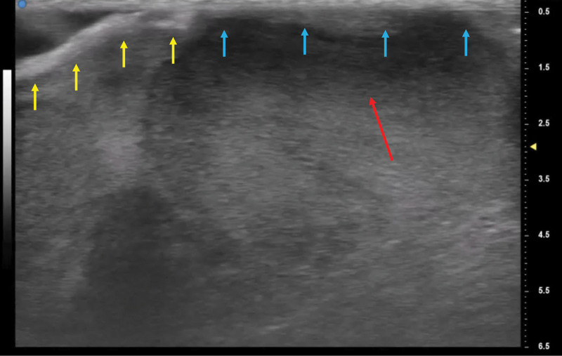
Aplasia Cutis Congenita (ACC) in the scalp is a rare congenital malformation. The treatment for ACC with large defects involving the scalp, bone, and the dura is challenging. Local debridement of necrotic tissue is important to prevent lethal complications such as infection and meningitis. However, debridement has the risk of damaging the sagittal sinus or the dura. Recent developments in ultra-high-frequency ultrasound(US) systems provide frequencies of 70 MHz and capability resolution as fine as 30 μm, which could allow precise imaging of small and thin anatomical structures. The study aimed to describe the methods of precise evaluation of the defect in the scalp and safe debridement using ultra-high-frequency US. This is the first report on direct observation of a newborn's brain using ultra-high-frequency US. The boy was delivered spontaneously with a large defect of the scalp and bone. After 14 days, due to signs of infection, local debridement was performed carefully under ultra-high-frequency US-based evaluation. The dura, the sagittal sinus, and the small anatomical structures such as arachnoid granulations could be observed. Because the brain herniation gradually aggravated, dural reconstruction using fascia lata and scalp reconstruction using transposition flap was performed. Finally, good skin coverage over the defects was obtained. This method minimizes the risk of damaging the sagittal sinus and the brain parenchyma, which may cause fatal complications. Although further clinical investigations will be required to confirm its efficacy, ultra-high-frequency US has the potential to be a useful device for ACC treatment.
Copyright © 2021 The Authors. Published by Wolters Kluwer Health, Inc. on behalf of The American Society of Plastic Surgeons.
Conflict of interest statement
Disclosure: The authors have no financial interest in relation to the content of this article.
Figures


Similar articles
-
Case report: Aplasia cutis congenita of the scalp with bone defect and an exposed sagittal sinus in a trisomy 13 newborn.Front Pediatr. 2023 Mar 31;11:1142950. doi: 10.3389/fped.2023.1142950. eCollection 2023. Front Pediatr. 2023. PMID: 37063682 Free PMC article.
-
Aplasia cutis congenita of the scalp, skull and dura associated with Adams-Oliver syndrome.Turk Neurosurg. 2008 Apr;18(2):191-3. Turk Neurosurg. 2008. PMID: 18597236
-
[Surgical management of aplasia cutis congenita].An Pediatr (Barc). 2015 Nov;83(5):341-5. doi: 10.1016/j.anpedi.2015.02.005. Epub 2015 Mar 21. An Pediatr (Barc). 2015. PMID: 25804551 Spanish.
-
Fatal superior sagittal sinus hemorrhage as a complication of aplasia cutis congenita: a case report and literature review.Forensic Sci Med Pathol. 2015 Jun;11(2):243-8. doi: 10.1007/s12024-014-9645-5. Epub 2015 Jan 23. Forensic Sci Med Pathol. 2015. PMID: 25614301 Review.
-
Scalp aplasia cutis congenita presenting with sagittal sinus hemorrhage.Arch Otolaryngol Head Neck Surg. 2001 Jan;127(1):71-4. doi: 10.1001/archotol.127.1.71. Arch Otolaryngol Head Neck Surg. 2001. PMID: 11177019 Review.
References
-
- Campbell W. Case of congenital ulcer on the cranium of a fetus, terminating in fatal hemorrhage, on 18th day after birth. J Med Sci (Edinburgh) 1826. 1826;(2):82–84.
-
- Schnabl SM, Horch RE, Ganslandt O, et al. . Aplasia cutis congenital—plastic reconstruction of three scalp and skull defects with two opposed scalp rotation flaps and split thickness skin grafting. Neuropediatrics. 2009;40:134–136. - PubMed
-
- Bajpai M, Pal K. Aplasia cutis cerebri with partial acrania—total reconstruction in a severe case and review of the literature. J Pediatr Surg. 2003;38:e4. - PubMed
-
- Ribuffo D, Costantini M, Gullo P, et al. . Aplasia cutis congenita of the scalp, the skull, and the dura. Scand J Plast Reconstr Surg Hand Surg. 2003;37:176–180. - PubMed
-
- Theile RJ, Lanigan MW, McDermant GR. Reconstruction of aplasia cutis congenita of the scalp by split rib cranioplasty and a free latissimus dorsi muscle flap in a nine month old infant. Br J Plast Surg. 1995;48:507–510. - PubMed
LinkOut - more resources
Full Text Sources