Review
. 2021 Oct;11(5):1140-1149.
doi: 10.21037/cdt-20-160.

Nutcracker syndrome: diagnosis and therapy

Affiliations
Review

Nutcracker syndrome: diagnosis and therapy

Marcin K Kolber et al. Cardiovasc Diagn Ther. 2021 Oct.

Abstract

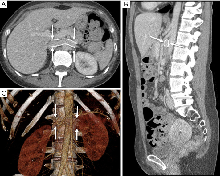
Nutcracker syndrome (NCS) is an extrinsic compression of the left renal vein (LRV) by the superior mesenteric artery (SMA) anteriorly and aorta posteriorly resulting in renal vascular congestion manifesting as hematuria, proteinuria, orthostatic hypotension, pain, or even renal dysfunction. Long-standing venous compression can encourage collateral drainage pathways through gonadal and pelvic veins, which may explain reported symptom and syndrome overlap with pelvic congestion syndrome. Diagnosis can be challenging and variable, frequently involving a combination of ultrasound Doppler, cross-sectional, and invasive imaging. Often, intravascular pressure measurements are required to prove a renocaval pressure gradient to aid in a definitive diagnosis. Conservative management is appropriate, especially in children, who tend to outgrow the disorder. In the interim, medical management with angiotensin converting enzyme inhibitors (ACEIs) is a useful therapy to manage orthostatic hypotension in the pediatric population. In adults, invasive therapies are more frequently pursued. These are aimed at relieving the extrinsic compression on the LRV. The standard of care is renal vein transposition, with renal autotransplantation reserved for recalcitrant cases. Endovascular stenting is a less invasive option. Laparoscopic placement of an exovascular stent is a newer therapy intended to minimize trauma to the LRV. In this review, we will discuss the clinical manifestations, diagnostic criterion, imaging features, and conservative and surgical therapies for this condition.

Keywords: Nutcracker syndrome (NCS); hematuria; left renal vein (LRV); vascular compression.


Conflict of interest statement

Conflicts of Interest: All authors have completed the ICMJE uniform disclosure form (available at http://dx.doi.org/10.21037/cdt-20-160). The series “Compressive Vascular Syndromes” was commissioned by the editorial office without any funding or sponsorship. Drs. MKK and SK served as the unpaid Guest Editors of the series. Dr. SK serves as an unpaid editorial board member of Cardiovascular Diagnosis and Therapy from May 2019 to Apr 2021. Dr. SK reports personal fees from Medtronic, personal fees from Dova Pharmaceuticals, personal fees from Koo Foundation, Taiwan, personal fees from GE healthcare, personal fees from Springer, personal fees from Elsevier, other from Althea Health Inc, CA, personal fees from Boston Scientific, personal fees from Penumbra, grants from BD, grants from NIH, outside the submitted work. The authors have no other conflicts of interest to declare.

Figures

Figure 1
Figure 1
Axial (A) and sagittal (B) venous-phase contrast-enhanced CTA demonstrating compression of the AM segment of the LRV (white arrow) between the SMA (dashed white arrow) and aorta (dotted black arrow). CTA, computed tomography angiography; AM, aortomesenteric; LRV, left renal vein; SMA, superior mesenteric artery.
Figure 2
Figure 2
Normal eft renal venography (without evidence of NCS), demonstrating minimal reflux into small-caliber lumbar (solid white arrow), gonadal (dashed white arrow), and adrenophrenic (dotted black arrow) veins. NCS, nutcracker syndrome.
Figure 3
Figure 3
Collateral venous drainage in NCS. (A) Left renal venography in NCS demonstrates poor opacification of the AM LRV (solid black arrows) and IVC (dotted black arrows) with preferential flow via the lumbar venous plexus (solid white arrows) and gonadal vein (dashed white arrows); (B) in females, an enlarged gonadal vein (dashed white arrows) can collateralize with an enlarged uterine venous plexus (dashed black arrows); (C) in males, left renal venous hypertension can contribute to varicocele (shown). NCS, nutcracker syndrome; AM, aortomesenteric; LRV, left renal vein; IVC, inferior vena cava.
Figure 4
Figure 4
DUS diagnosis of NCS. (A) Grayscale ultrasound identifies the compressed AM segment LRV (solid white arrows) and more peripherally, relative dilation of the more peripheral vein (dashed arrows); (B) DUS at the hilar LRV with low peak velocity (20.5 cm/sec); (C) similar measurements at the aorto-mesenteric segment with high peak velocity (159 cm/sec), yielding a ratio of 8:1. DUS, Doppler ultrasound; NCS, nutcracker syndrome; AM, aortomesenteric; LRV, left renal vein.
Figure 5
Figure 5
CT diagnosis of NCS. (A) CTA demonstrates an aorta-to-SMA angle of <35° in sagittal plane; (B) comparison measurements of aorto-mesenteric (2.3 mm) to hilar (12.5 mm) LRV in the same patient yields a “compression ratio” of 5.4. CT, computed tomography; NCS, nutcracker syndrome; CTA, computed tomography angiography; SMA, superior mesenteric artery; LRV, left renal vein.
Figure 6
Figure 6
MRI findings in NCS. Axial balanced steady-state gradient echo (A), axial T1 post-contrast mDIXON (B), and obliqued sagittal T1 post-contrast mDIXON (C) sequences demonstrate classic findings of LRV compression with “beak sign” (white arrows) at the aortic margin (MRIs performed in different patients). MRI, magnetic resonance imaging; NCS, nutcracker syndrome; LRV, left renal vein.
Figure 7
Figure 7
Left renal venography and simultaneous invasive pressure measurements via the vascular sheath in the IVC (dashed white arrows) and catheter in the hilar LRV (solid red arrows). Screenshot of the pressure monitor shows venous pressure waveforms (corresponding arrows) and pressure measurements with mean sheath pressure 0 mmHg and mean left renal catheter pressure 4 mmHg (gradient =4 mmHg), diagnostic of NCS. Note a vascular plug (dotted black arrow) and coils (dashed black arrows) in an embolized gonadal vein (common treatment for associated pelvic congestion syndrome). IVC, inferior vena cava; LRV, left renal vein; NCS, nutcracker syndrome.
Figure 8
Figure 8
Sagittal (A) and axial (B) CTA following laparoscopic exovascular stent (arrows) placement in a patient with previously diagnosed NCS. CTA, computed tomography angiography; NCS, nutcracker syndrome.
Figure 9
Figure 9
Axial (A), sagittal (B), and 3D-reconstruction (C) CTA following endovascular stent (arrows) deployment in a patient with previously diagnosed NCS. CTA, computed tomography angiography; NCS, nutcracker syndrome.

References

    1. de Schepper A. "Nutcracker" phenomenon of the renal vein and venous pathology of the left kidney. J Belge Radiol 1972;55:507-11. - PubMed
    1. Kim KW, Cho JY, Kim SH, et al. Diagnostic value of computed tomographic findings of nutcracker syndrome: correlation with renal venography and renocaval pressure gradients. Eur J Radiol 2011;80:648-54. 10.1016/j.ejrad.2010.08.044 - DOI - PubMed
    1. Ananthan K, Onida S, Davies AH. Nutcracker syndrome: an update on current diagnostic criteria and management guidelines. Eur J Vasc Endovasc Surg 2017;53:886-94. 10.1016/j.ejvs.2017.02.015 - DOI - PubMed
    1. He Y, Wu Z, Chen S, et al. Nutcracker syndrome--how well do we know it? Urology 2014;83:12-7. 10.1016/j.urology.2013.08.033 - DOI - PubMed
    1. Shin JI, Lee JS, Kim MJ. The prevalence, physical characteristics and diagnosis of nutcracker syndrome. Eur J Vasc Endovasc Surg 2006;32:335-6. 10.1016/j.ejvs.2006.04.030 - DOI - PubMed