

Cytologic Categorization with Risk Stratification of Endoscopic Ultrasound-Guided Fine Needle Aspiration from Pancreatic Lesions Based on Guidelines of the Papanicolaou Society of Cytopathology: 12-Year Tertiary Care Experience
- PMID: 34816002
- PMCID: PMC8605790
- DOI: 10.15190/d.2021.13
Cytologic Categorization with Risk Stratification of Endoscopic Ultrasound-Guided Fine Needle Aspiration from Pancreatic Lesions Based on Guidelines of the Papanicolaou Society of Cytopathology: 12-Year Tertiary Care Experience
Abstract
Background and aims: Pancreatic malignancy is an important cause of cancer mortality worldwide. Endoscopic ultrasound-guided fine-needle aspiration (EUS-FNA) plays a crucial role in the pre-operative diagnosis of pancreatic lesions. In this study, we have analyzed the cytological spectrum of pancreatic lesions in the Indian population over 12 years, categorized them according to the Papanicolaou Society of Cytopathology System for Reporting Pancreaticobiliary Cytology (PSCPC), and assessed the risk of malignancy (ROM) for each of the categories.
Methods: A computerized data search from January 2008 to December 2019 revealed 581 pancreatic EUS-FNA samples, among which surgical follow-up was available for 73 cases. All cytological specimens were reviewed and prospectively classified into one of the six diagnostic categories proposed by the PSCPC. Subsequently, a cytohistological correlation was performed and the ROM was calculated for each category.
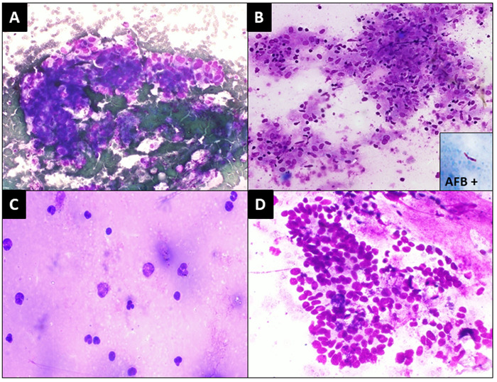
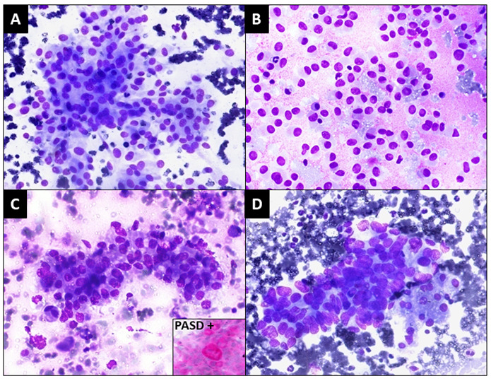
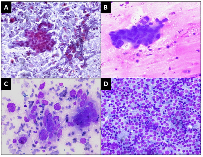
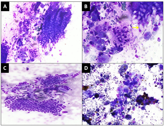
Results: The cytologic diagnoses included 50 nondiagnostic (category I), 175 negative for malignancy (category II), 19 atypical (category III), 27 neoplastic:benign (category IVA), 30 neoplastic:other (category IVB), 26 suspicious (category V), and 254 malignant (category VI) cases. ROM for non-diagnostic aspirates, nonneoplastic benign specimens, atypical cases, neoplastic:benign, neoplastic:other, suspicious for malignancy, and the malignant category was 16.7%, 7.1%, 33.3%, 0.0%, 20.0%, 100%, and 78.6%, respectively.
Conclusion: We document an increased risk of malignancy from category I to category VI of the PSCPC. The malignancy risk for category VI (malignant) was statistically significant in our study but was lower in comparison to the values reported by other authors. Nonetheless, such an approach would establish transparent communication between the pathologist and the clinician, as well as aid the clinician in decision making, particularly in intermediate categories.
Keywords: EUS-FNA; Papanicolaou Society of Cytopathology; pancreas; risk of malignancy; risk stratification..
Copyright © 2021, Nishith N et al., Applied Systems and Discoveries Journals.
Conflict of interest statement
Conflict of interests: The authors declare no competing interests.
Figures
Similar articles
-
Update on risk stratification in the Papanicolaou Society of Cytopathology System for Reporting Pancreaticobiliary Cytology categories: 3-Year, prospective, single-institution experience.Cancer Cytopathol. 2020 Jan;128(1):29-35. doi: 10.1002/cncy.22199. Epub 2019 Nov 13. Cancer Cytopathol. 2020. PMID: 31722125
-
Risk of malignancy associated with diagnostic categories of the proposed World Health Organization International System for Reporting Pancreaticobiliary Cytopathology.Cancer Cytopathol. 2022 Mar;130(3):195-201. doi: 10.1002/cncy.22514. Epub 2021 Oct 8. Cancer Cytopathol. 2022. PMID: 34623767
-
Malignancy risk associated with diagnostic categories defined by the Papanicolaou Society of Cytopathology pancreaticobiliary guidelines.Cancer Cytopathol. 2014 Jun;122(6):420-7. doi: 10.1002/cncy.21386. Epub 2013 Dec 11. Cancer Cytopathol. 2014. PMID: 24339321
-
The frequency and cancer risk associated with the atypical cytologic diagnostic category in endoscopic ultrasound-guided fine-needle aspiration specimens of solid pancreatic lesions: a meta-analysis and argument for a Bethesda System for Reporting Cytopathology of the Pancreas.Cancer Cytopathol. 2013 Nov;121(11):620-8. doi: 10.1002/cncy.21337. Epub 2013 Jul 23. Cancer Cytopathol. 2013. PMID: 23881871 Review.
-
Diagnostic Performance of Pancreatic Cytology with the Papanicolaou Society of Cytopathology System: A Systematic Review, before Shifting into the Upcoming WHO International System.Int J Mol Sci. 2022 Jan 31;23(3):1650. doi: 10.3390/ijms23031650. Int J Mol Sci. 2022. PMID: 35163571 Free PMC article.
Cited by
-
Reporting Pancreatic FNAC using the Papanicolaou System: Still a Diagnostic Challenge.J Cytol. 2024 Apr-Jun;41(2):123-130. doi: 10.4103/joc.joc_90_23. Epub 2024 Apr 24. J Cytol. 2024. PMID: 38779600 Free PMC article.
-
The World Health Organization System for Reporting Pancreaticobiliary Cytopathology: Standardized Categories and Practical Approaches to Pancreatic Lesions.J Clin Transl Pathol. 2024 Sep;4(3):122-135. doi: 10.14218/jctp.2024.00034. Epub 2024 Sep 25. J Clin Transl Pathol. 2024. PMID: 40191142 Free PMC article.
-
Intraoperative Tumor Detection Using Pafolacianine.Int J Mol Sci. 2022 Oct 25;23(21):12842. doi: 10.3390/ijms232112842. Int J Mol Sci. 2022. PMID: 36361630 Free PMC article. Review.
-
Echoendoscopic ultrasound pancreatic adenocarcinoma diagnosis and theranostic approach: should KRAS mutation research be recommended in everyday practice?Therap Adv Gastroenterol. 2024 Jan 17;17:17562848231224943. doi: 10.1177/17562848231224943. eCollection 2024. Therap Adv Gastroenterol. 2024. PMID: 38250014 Free PMC article.
References
-
- Global cancer statistics 2018: GLOBOCAN estimates of incidence and mortality worldwide for 36 cancers in 185 countries. Bray Freddie, Ferlay Jacques, Soerjomataram Isabelle, Siegel Rebecca L, Torre Lindsey A, Jemal Ahmedin. CA: a cancer journal for clinicians. 2018;68(6):394–424. - PubMed
-
- Standardized terminology and nomenclature for pancreatobiliary cytology: the Papanicolaou Society of Cytopathology guidelines. Pitman Martha B, Centeno Barbara A, Ali Syed Z, Genevay Muriel, Stelow Ed, Mino-Kenudson Mari, Fernandez-del Castillo Carlos, Max Schmidt C, Brugge William, Layfield Lester. Diagnostic cytopathology. 2014;42(4):338–50. - PubMed
-
- Standardized terminology and nomenclature for pancreatobiliary cytology: The Papanicolaou Society of Cytopathology Guidelines. Pitman Martha B, Centeno Barbara A, Ali Syed Z, Genevay Muriel, Stelow Ed, Mino-Kenudson Mari, Castillo Carlos Fernandez-Del, Schmidt C Max, Brugge William R, Layfield Lester J. CytoJournal. 2014;11(Suppl 1):3. - PMC - PubMed
LinkOut - more resources
Full Text Sources