Review
. 2022 Jan;37(1):6-15.
doi: 10.1002/mds.28858. Epub 2021 Nov 23.

Parkinson's Disease Modification Through Abl Kinase Inhibition: An Opportunity

Affiliations
Review

Parkinson's Disease Modification Through Abl Kinase Inhibition: An Opportunity

Milton H Werner et al. Mov Disord. 2022 Jan.

Abstract

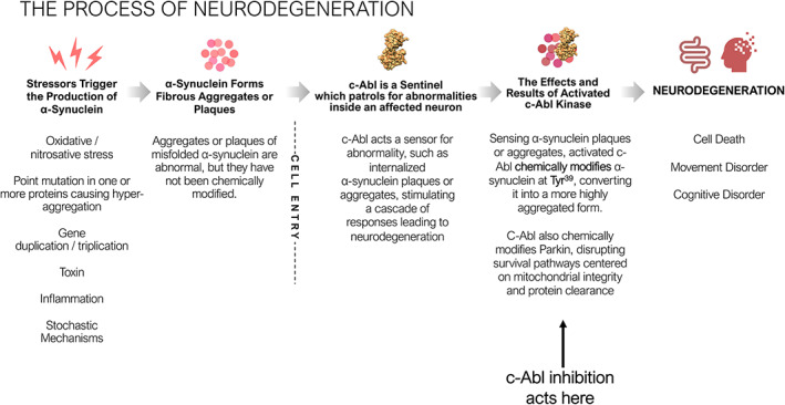
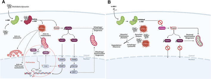
Parkinson's disease (PD) is the second most prevalent neurodegenerative disease of the central nervous system, with an estimated 5 000 000 cases worldwide. Historically characterized by the progressive loss of dopaminergic neurons in the substantia nigra pars compacta, PD pathology is now known to be widespread and to affect serotonin, cholinergic and norepinephrine neurons as well as nerve cells in the olfactory system, cerebral hemisphere, brain stem, spinal cord, and peripheral autonomic nervous system. PD pathology is characterized by the accumulation of misfolded α-synuclein, which is thought to play a critical role in the etiopathogenesis of the disease. Animal models of PD suggest that activation of the Abelson tyrosine kinase (c-Abl) plays an essential role in the initiation and progression of α-synuclein pathology and neurodegeneration. These studies demonstrate that internalization of misfolded α-synuclein activates c-Abl, which phosphorylates α-synuclein and promotes α-synuclein pathology within the affected neurons. Additionally, c-Abl inactivates parkin, disrupting mitochondrial quality control and biogenesis, promoting neurodegeneration. Post-mortem studies of PD patients demonstrate increased levels of tyrosine phosphorylated α-synuclein, consistent with the activation of c-Abl in human disease. Although the c-Abl inhibitor nilotinib failed to demonstrate clinical benefit in two double-blind trials, novel c-Abl inhibitors have been developed that accumulate in the brain and may inhibit c-Abl at saturating levels. These novel inhibitors have demonstrated benefits in animal models of PD and have now entered clinical development. Here, we review the role of c-Abl in the neurodegenerative disease process and consider the translational potential of c-Abl inhibitors from model studies to disease-modifying therapies for Parkinson's disease. © 2021 Inhibikase Therapeutics, Inc. Movement Disorders published by Wiley Periodicals LLC on behalf of International Parkinson Movement Disorder Society.

Keywords: Abelson tyrosine kinase; Parkinson's disease; disease-modification.


Figures

FIG 1
FIG 1
Feature map of α‐synuclein, c‐Abl, and parkin. (A) α‐Synuclein is a small, irregularly structured protein of 140 amino acids, which is comprised of a 61 amino acid amphipathic helical bundle where both the c‐Abl phosphorylation site at Tyr39 (Y39) and one of six disease associated point mutants (A53T) is located and associated with inherited Parkinson's disease. The non‐amyloid‐β‐component (NAC) is followed by an irregularly structured acidic region where both Tyr125 and Ser129 reside, whereas Tyr39 resides in the amphipathic coiled‐coil region. Above these chemical modification sites are the putative kinases implicated in phosphorylation of these sites. (B) c‐Abl is a large, multidomain protein of 1142 amino acids, wherein the regulatory regions of the myristylation cap, SH3, and SH2 domains reside in the N‐terminal half of the protein, followed by tyrosine kinase domain. The kinase core of the c‐Abl protein has a domain organization similar to that of the Src family kinases, with sequential Src homology (SH) 3 and SH2 domains, an SH2/kinase linker, and a bi‐lobed kinase domain. This core is flanked by an N‐terminal “cap” (N‐cap) region with a signal sequence for myristylation, which serves dual roles in regulation of kinase activity and in membrane localization. C‐terminal to the kinase domain is a long region of >600 amino acids encoded by a single exon, which controls interaction of Abl with other SH3‐containing proteins and the actin cytoskeleton. This region also regulates nuclear‐cytoplasmic shuttling of the kinase. (C) Parkin is a protein of 465 amino acids consisting of a Ubiquitin‐like domain (Ubl) at its N terminus and four zinc‐coordinating RING‐like domains: RING0, RING1, in‐between RING (IBR) and RING2, and a repressor element of parkin (REP) domain. It is classified as a RING‐between‐RING or RBR E3 ubiquitin ligase. More than 120 pathogenic PD mutations are spread throughout parkin domains, attesting to critical functions for each of the individual domain. The Ubl domain is involved in substrate recognition, binding SH3 and ubiquitin interacting motif (UIM) domains, proteasome association, and regulation of cellular parkin levels and activity.
FIG 2
FIG 2
The Process of Neurodegeneration. Environmental, genetic and biochemical triggers lead to α‐synuclein aggregate formation. These aggregates are abnormal, may have diverse quaternary structures and are believed to be benign. The α‐synuclein aggregates or plaques that matter arise after the affected neurons take up the benign aggregates or plaques. Once internalized, c‐Abl, a sentinel capable of recognizing abnormalities, is activated, leading to chemical modification by phosphorylation of the internalized aggregates or plaques at Tyr39. c‐Abl also inactivates the ubiquitin E3 ligase Parkin, which drives neurodegeneration through a combination of mitophagy and/or parthanatos and shuts off pathways for clearance of phosphorylated and/or internalized α‐synuclein aggregates or plaques.
FIG 3
FIG 3
The biochemistry of Parkinson's disease initiation and progression and how to disrupt it. (A) The process of neurodegeneration. Misfolded α‐synuclein can arise from a variety of factors (see text). Misfolded α‐synuclein may form within the neuron or by transfer through cell surface receptors or by crossing membrane bilayers. Within a neuron, misfolded α‐synuclein is ‘sensed’ and c‐Abl activated, driving the formation of pathologic α‐synuclein by chemical modification (p‐Syn). Chemical modification creates a form of α‐synuclein that represents the pathologic species of the disease leading to disruption of mitochondrial integrity, negatively impact the endosome, disrupt nucleosomal structure and modulate transcription of certain genes., , , , , , , , , , , , , , , , C‐Abl also inactivates parkin by chemical modification, which affects mitochondrial quality control and suppresses protein clearance mechanisms., , , , , , , , , Parkin inactivation suppresses the complex interplay between parkin and pink1 at the mitochondrion, which act in concert to maintain mitochondrial integrity, quality and regulate mitochondrial biogenesis. Parkin inactivation leads to the accumulation of toxic parkin substrates, such as the parkin interacting substrate (PARIS), the aminoacyl tRNA synthetase complex‐interacting multifunctional protein 2 (AIMP2) and the far upstream element‐binding protein 1 (FBP1)., , , , , , , , , PARIS and AIMP2 accumulate in adult conditional parkin knockout mice and MPTP‐intoxicated mice as well as in patients with PD. Increased levels of PARIS can lead to mitochondrial dysfunction through downregulation of the peroxisome proliferator‐activated receptor‐gamma coactivator (PGC)‐1α transcriptional co‐activator protein and loss of DA neurons in a PARIS‐dependent manner. PGC1‐α is a transcriptional co‐activator that plays a central role in regulation of energy metabolism. Overexpression of AIMP2 leads to an age dependent, selective degeneration of DA neurons through activation of poly (ADP‐ribose) polymerase 1 (PARP1), driving PARP1‐mediated parthanatos. This suggests that PARIS and AIMP2 may be important contributors to the loss of DA neurons and possibly other vulnerable neurons following parkin inactivation. Inactivation of parkin also disrupts protein clearance mechanisms through autophagy, lysosomal, and proteasomal degradation pathways. (B) The consequences of c‐Abl inhibitor treatment on the process of neurodegenerative disease. Inhibition of c‐Abl precludes c‐Abl activation, blocking the build‐up of toxic parkin substrates PARIS and AIMP2 and terminating downstream events. This also re‐establishes normal mitochondrial quality control and biogenesis. Model studies demonstrate that modified and unmodified α‐synuclein aggregates are shunted to lysosomal or proteasomal degradation pathways for clearance with concomitant recovery of motor function.

References

    1. Bach JP, Ziegler U, Deuschl G, et al. Projected numbers of people with movement disorders in the years 2030 and 2050. Mov Disord 2011;26:2286–2290. - PubMed
    1. Olanow CW, Stern MB, Sethi K. Scientific and clinical basis for the treatment of PD – 2009. Neurology 2009;72(21 suppl 4):S1–S136. - PubMed
    1. Jellinger KA. Neuropathology of sporadic Parkinson's disease: evaluation and changes of concepts. Mov Disord 2012;27:8–30. - PubMed
    1. Schapira AHV, Chaudhuri KR, Jenner P. Non‐motor features of Parkinson disease. Nat Rev Neurosci 2017;18:435–450. - PubMed
    1. Lindholm D, Pham DD, Cascone A, Eriksson O, Wennerberg K, Saarma M. c‐Abl inhibitors enable insights into the pathophysiology and neuroprotection in Parkinson's disease. Front Aging Neurosci 2016;8:254 - PMC - PubMed

Publication types

MeSH terms

Substances