. 2022 Jan;9(3):e2102460.
doi: 10.1002/advs.202102460. Epub 2021 Nov 23.

Fine Particulate Matter Induces Childhood Asthma Attacks via Extracellular Vesicle-Packaged Let-7i-5p-Mediated Modulation of the MAPK Signaling Pathway

Affiliations

Fine Particulate Matter Induces Childhood Asthma Attacks via Extracellular Vesicle-Packaged Let-7i-5p-Mediated Modulation of the MAPK Signaling Pathway

Rui Zheng et al. Adv Sci (Weinh). 2022 Jan.

Abstract

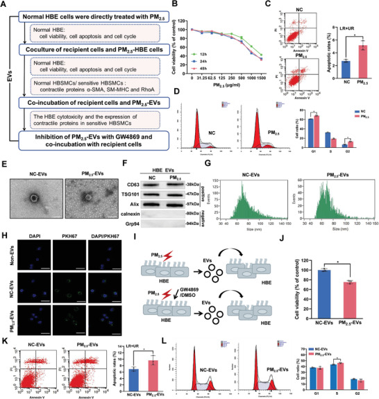
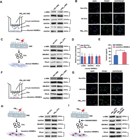
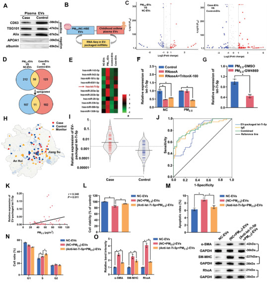
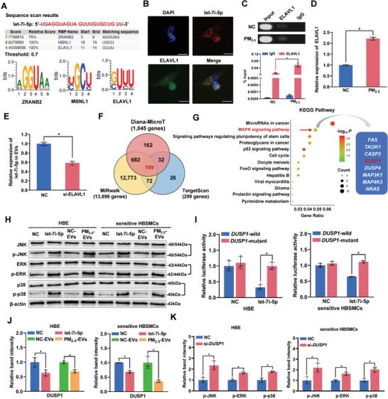
Fine particulate matter less than 2.5 µm in diameter (PM2.5 ) is a major risk factor for acute asthma attacks in children. However, the biological mechanism underlying this association remains unclear. In the present study, PM2.5 -treated HBE cells-secreted extracellular vesicles (PM2.5 -EVs) caused cytotoxicity in "horizontal" HBE cells and increased the contractility of "longitudinal" sensitive human bronchial smooth muscle cells (HBSMCs). RNA sequencing showed that let-7i-5p is significantly overexpressed in PM2.5 -EVs and asthmatic plasma; additionally, its level is correlated with PM2.5 exposure in children with asthma. The combination of EV-packaged let-7i-5p and the traditional clinical biomarker IgE exhibits the best diagnostic performance (area under the curve [AUC] = 0.855, 95% CI = 0.786-0.923). Mechanistically, let-7i-5p is packaged into PM2.5 -EVs by interacting with ELAVL1 and internalized by both "horizontal" recipient HBE cells and "longitudinal" recipient-sensitive HBSMCs, with subsequent activation of the MAPK signaling pathway via suppression of its target DUSP1. Furthermore, an injection of EV-packaged let-7i-5p into PM2.5 -treated juvenile mice aggravated asthma symptoms. This comprehensive study deciphered the remodeling of the extracellular environment mediated by the secretion of let-7i-5p-enriched EVs during PM2.5 -induced asthma attacks and identified plasma EV-packaged let-7i-5p as a novel predictor of childhood asthma.

Keywords: MAPK signaling pathway; PM2.5; childhood asthma; extracellular vesicles; let-7i-5p.


Conflict of interest statement

The authors declare no conflict of interest.

Figures

Figure 1
Figure 1
The effect of PM2.5‐EVs on cytotoxicity in HBE cells. PM2.5‐treated HBE cells were designated PM2.5‐HBE cells. EVs isolated from PM2.5‐treated HBE cells and NC‐treated HBE cells were designated PM2.5‐EVs and NC‐EVs, respectively. A) Flowchart of the strategy used to evaluate the effect of PM2.5‐EVs on cytotoxicity in HBE cells and the contractility of sensitive HBSMCs. B) The viability of HBE cells was evaluated using a CCK‐8 assay after PM2.5 treatment. C) The apoptosis of HBE cells was assessed using flow cytometry after PM2.5 treatment. D) The cell cycle of HBE cells was analyzed using flow cytometry after PM2.5 treatment. E) Purified PM2.5/NC‐EVs were identified using TEM. Scale bar, 100 nm. F) The EV‐specific markers (CD63, TSG101 and Alix) and negative controls (calnexin and Grp94) were evaluated using Western blotting. G) The size distributions of PM2.5/NC‐EVs were confirmed using NanoFCM. H) Representative fluorescence images of HBE cells after an incubation with PM2.5/NC‐EVs labelled with PKH67 (green) or non‐EVs. Scale bar, 25 µm. I) Schematic illustration of HBE cells incubated with PM2.5‐EVs or PM2.5‐EVs pretreated with DMSO/GW4869. J) The viability of HBE cells was evaluated using a CCK‐8 assay after an incubation with PM2.5/NC‐EVs. K) The apoptosis of HBE cells was assessed using flow cytometry after an incubation with PM2.5/NC‐EVs. L) The cell cycle of HBE cells was analyzed using flow cytometry after an incubation with PM2.5/NC‐EVs. Statistical significance was assessed using two‐tailed Student's t‐test. Values represent means ± SD. * p < 0.05.
Figure 2
Figure 2
Effect of PM2.5‐EVs on the contractility of sensitive HBSMCs. The PM2.5/NC‐treated HBE cells are designated PM2.5/NC‐HBE cells. The red lightning signal represents PM2.5 exposure. A) The levels of the contractile proteins α‐SMA, SM‐MHC and RhoA in normal HBSMCs cocultured with PM2.5/NC‐HBE cells were analyzed using Western blotting. B) Representative fluorescence images of normal HBSMCs after an incubation with PM2.5/NC‐EVs labelled with PKH67 (green) or non‐EVs. Scale bar, 25 µm. C) The levels of the contractile proteins α‐SMA, SM‐MHC and RhoA in normal HBSMCs incubated with PM2.5/NC‐EVs were analyzed using Western blotting. D) The band intensity of contractile proteins in normal HBSMCs incubated with PM2.5/NC‐EVs. E) Normal HBSMCs were treated with IL‐13 to establish a sensitive HBSMC model. The expression of eotaxin in normal and sensitive HBSMCs was measured using an enzyme‐linked immunosorbent assay (ELISA) kit. F) The levels of the contractile proteins α‐SMA, SM‐MHC and RhoA in sensitive HBSMCs cocultured with PM2.5/NC‐HBE cells were analyzed using Western blotting. G) Representative fluorescence images of sensitive HBSMCs after an incubation with PM2.5/NC‐EVs labelled with PKH67 (green) or non‐EVs. Scale bar, 25 µm. H) The contractile proteins α‐SMA, SM‐MHC and RhoA in sensitive HBSMCs incubated with PM2.5/NC‐EVs were analyzed using Western blotting. I) The contractile proteins α‐SMA, SM‐MHC and RhoA in sensitive HBSMCs incubated with PM2.5/NC‐EVs pretreated with DMSO/GW4869 were analyzed using Western blotting. Statistical significance was assessed using two‐tailed Student's t‐test. Values represent means ± SD. * p < 0.05.
Figure 3
Figure 3
The role of EV‐packaged let‐7i‐5p in children with asthma and the phenotype of recipient cells. HBE cells were transfected with NC‐ or let‐7i‐5p inhibitor, exposed to NC/PM2.5 and then isolated the corresponding EVs, namely, NC‐EVs, (NC+PM2.5)‐EVs, and (Anti‐let‐7i‐5p+PM2.5)‐EVs, respectively. A) EV‐specific markers (CD63, TSG101 and Alix) and negative controls (APOA1 and albumin) were evaluated using Western blotting. B) Schematic model showing the generation of EV‐packaged miRNA expression profiles. C) Volcano plots of different EV‐packaged miRNAs in PM2.5‐treated HBE cell models and children with asthma. The red and blue plots indicate the differentially expressed EV‐packaged miRNAs. D) Venn diagram showing the overlapping upregulated EV‐packaged miRNAs. E) Hierarchical clustering analysis of the 11 overlapping upregulated EV‐packaged miRNAs identified from the Venn diagram. F) The level of let‐7i‐5p in the culture medium of PM2.5/NC‐treated HBE cells incubated with 2 µg mL−1 RNaseA alone or in combination with 0.1% Triton X‐100 was determined using RT‐qPCR. G) The level of let‐7i‐5p in the culture medium of PM2.5‐treated HBE cells exposed to DMSO or GW4869 was determined using RT‐qPCR. H) Locations of the individuals’ residences and air quality monitoring stations. I) The level of plasma EV‐packaged let‐7i‐5p was determined using RT‐qPCR. J) ROC curve analysis of plasma levels of EV‐packaged let‐7i‐5p and IgE. K) Correlation analysis was performed between plasma EV‐packaged let‐7i‐5p and PM2.5 exposure levels in children with asthma. L) The viability of HBE cells incubated with NC‐EVs, (NC+PM2.5)‐EVs or (Anti‐let‐7i‐5p+PM2.5)‐EVs was evaluated using a CCK‐8 assay. M) The apoptosis of HBE cells incubated with NC‐EVs, (NC+PM2.5)‐EVs or (Anti‐let‐7i‐5p+PM2.5)‐EVs was assessed using flow cytometry. N) The cell cycle of HBE cells incubated with NC‐EVs, (NC+PM2.5)‐EVs, or (Anti‐let‐7i‐5p+PM2.5)‐EVs was analyzed using flow cytometry. O) The levels of contractile proteins in sensitive HBSMCs incubated with NC‐EVs, (NC+PM2.5)‐EVs, or (Anti‐let‐7i‐5p+PM2.5)‐EVs were measured using Western blotting. Statistical significance was assessed using two‐tailed Student's t‐test. Values represent means ± SD. * p < 0.05.
Figure 4
Figure 4
Let‐7i‐5p is packaged into PM2.5‐EVs in an ELAVL1‐dependent manner and regulates the DUSP1 expression level in recipient cells. A) The specific interaction between the let‐7i‐5p sequence and RNA‐binding protein motifs was predicted by RBPDB (threshold value = 0.7). B) Immunofluorescence assessment of let‐7i‐5p (red) and ELAVL1 (green) colocalization in HBE cells. Scale bar, 20 µm. C) Upper panel: image of gel electrophoresis of PCR products from the RIP assay. Lower panel: an RIP assay with an anti‐ELAVL1 antibody or control IgG was performed on PM2.5/NC‐treated HBE cells. The level of let‐7i‐5p in the immunoprecipitates was determined using RT‐qPCR. D) The level of the ELAVL1 mRNA in PM2.5/NC‐treated HBE cells was determined using RT‐qPCR. E) The level of let‐7i‐5p in EVs derived from HBE cells transfected with si‐ELAVL1 was determined using RT‐qPCR. F) The potential target genes containing the let‐7i‐5p seed sequence were predicted using the Diana‐MicroT, MiRwalk and TargetScan databases. G) KEGG pathway enrichment analysis of the 169 target genes. H) The protein levels of MAPK pathway‐related signaling molecules in NC mimic‐, let‐7i‐5p mimic‐, NC‐EVs‐, or PM2.5‐EVs‐treated recipient cells (HBE cells and sensitive HBSMCs) were determined using Western blotting. I) The binding affinity of let‐7i‐5p for DUSP1 was assessed using a dual‐luciferase reporter assay. J) The expression of the DUSP1 protein in NC mimic‐, let‐7i‐5p mimic‐, NC‐EVs, or PM2.5‐EVs‐treated HBE cells (left panel) and sensitive HBSMCs (right panel) was detected using Western blotting. K) The protein levels of MAPK pathway‐related signaling molecules in NC‐ or si‐DUPS1‐transfected HBE cells (left panel) and sensitive HBSMCs (right panel) were determined using Western blotting. Statistical significance was assessed using a two‐tailed Student's t‐test. Values represent means ± SD. * p < 0.05.
Figure 5
Figure 5
EV‐packaged let‐7i‐5p induces asthma in vivo. Thirty‐two female BALB/c mice were randomly assigned to the OVA group (group 1), OVA+PM2.5 group (group 2), OVA+PM2.5+NC‐EVs group (group 3), or OVA+PM2.5+let‐7i‐5p‐EVs group (group 4). A) Schematic showing the process used to establish the mouse model. The green arrows indicate that each mouse was injected with 28.8 µL of the OVA suspension containing 3.6 µg of OVA and 144 µg of Al(OH)3. The yellow arrows indicate that mice were exposed to 2% OVA for 20 min. The grey arrows indicate that mice were exposed to 1.57 mg kg−1 PM2.5. The blue arrows indicate that mice were challenged by an endotracheal instillation of 2 µg of NC‐EVs. The red arrows indicate that mice were challenged by an endotracheal instillation of 2 µg of let‐7i‐5p‐EVs. B) Detection of bronchoconstriction using myography. C) The level of EV‐packaged let‐7i‐5p in BALF was determined using RT‐qPCR. D) The level of DUSP1 in BALF was determined using RT‐qPCR. E) Representative images of H&E staining in lung tissues and total lung injury scores for each group. Scale bar, 50 µm. F) Representative photographs and quantification of DUSP1 immunostaining in lung tissues. Scale bar, 50 µm. G) The level of let‐7i‐5p in lung tissues was determined using RT‐qPCR. H) The concentration of IL‐6 in BALF was measured using ELISA. I) In the proposed model, EV‐packaged let‐7i‐5p secreted by PM2.5‐treated HBE cells induce asthma by regulating the expression of its target gene DUSP1 and activating the MAPK signaling pathway in recipient cells. MVB, multivesicular bodies. Statistical significance was assessed using two‐tailed Student's t‐test. Values represent means ± SD. * p < 0.05.

Similar articles

Cited by

References

    1. Hekking P. W., Wener R. R., Amelink M., Zwinderman A. H., Bouvy M. L., Bel E. H., J. Allergy Clin. Immunol. 2015, 135, 896. - PubMed
    1. Hallas H. W., Chawes B. L., Rasmussen M. A., Arianto L., Stokholm J., Bønnelykke K., Bisgaard H., PLoS Med. 2019, 16, e1002722. - PMC - PubMed
    1. Lancet Respir. Med. 2017, 5, 691. - PubMed
    1. Lancet 2018, 392, 1736. - PubMed
    1. Papi A., Brightling C., Pedersen S. E., Reddel H. K., Lancet 2018, 391, 783. - PubMed

Publication types