. 2022 Feb 9;96(3):e0162621.
doi: 10.1128/JVI.01626-21. Epub 2021 Nov 24.

Analysis of Glycosylation and Disulfide Bonding of Wild-Type SARS-CoV-2 Spike Glycoprotein

Affiliations

Analysis of Glycosylation and Disulfide Bonding of Wild-Type SARS-CoV-2 Spike Glycoprotein

Shijian Zhang et al. J Virol. .

Abstract

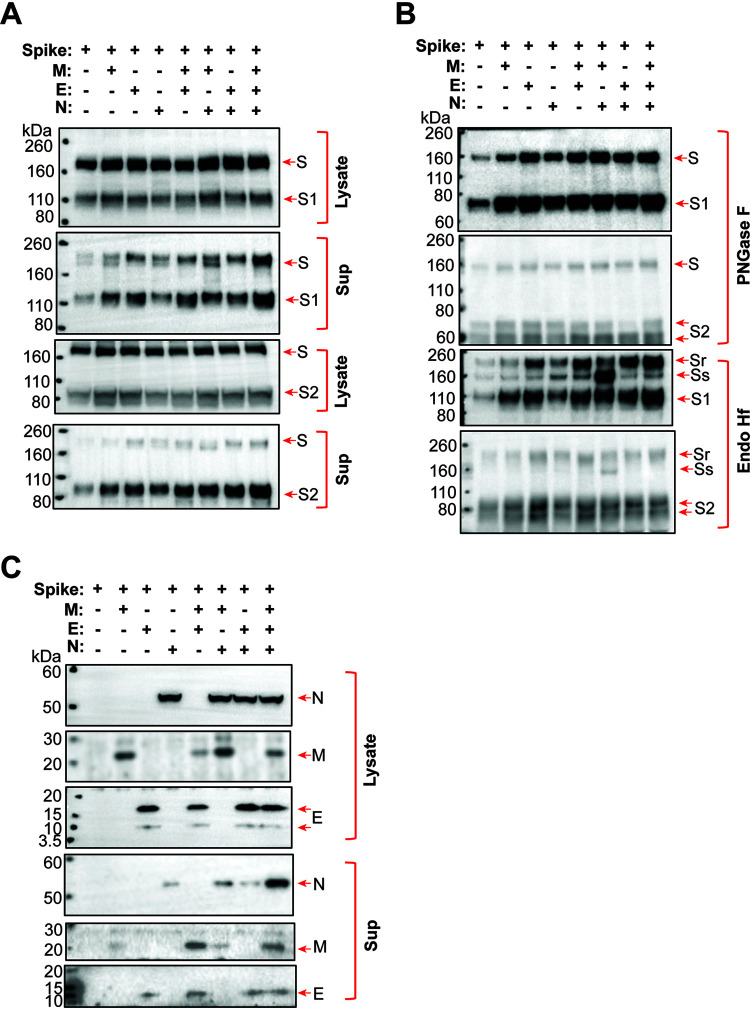
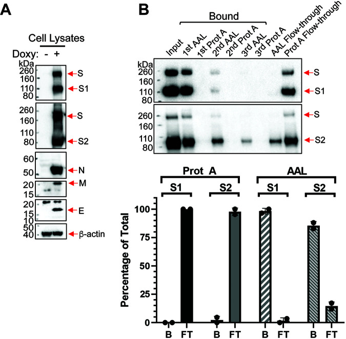
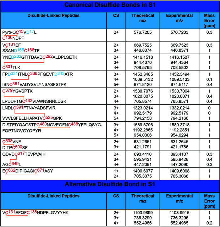
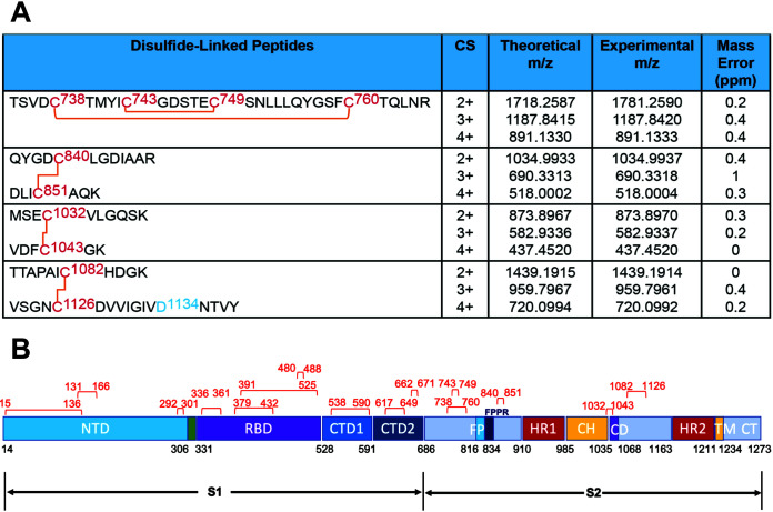
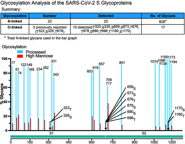
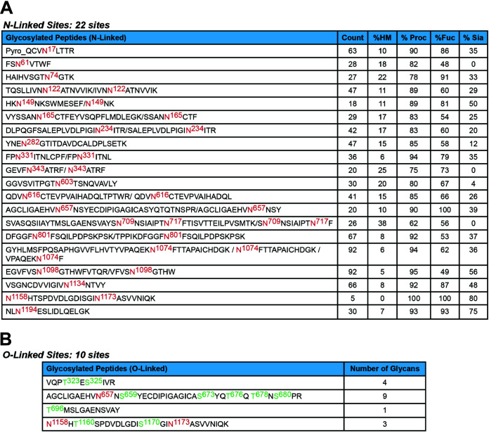
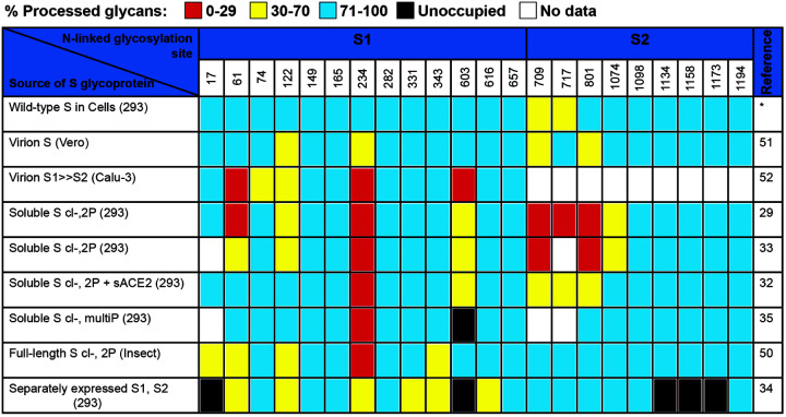
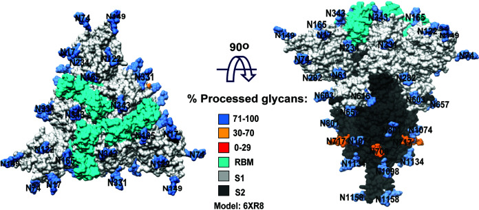
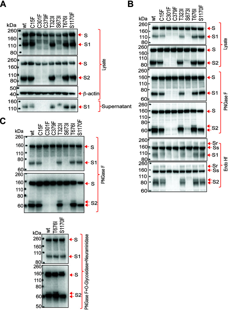
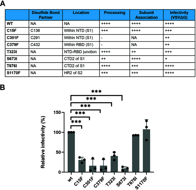
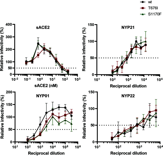
The SARS-CoV-2 coronavirus, the etiologic agent of COVID-19, uses its spike (S) glycoprotein anchored in the viral membrane to enter host cells. The S glycoprotein is the major target for neutralizing antibodies elicited by natural infection and by vaccines. Approximately 35% of the SARS-CoV-2 S glycoprotein consists of carbohydrate, which can influence virus infectivity and susceptibility to antibody inhibition. We found that virus-like particles produced by coexpression of SARS-CoV-2 S, M, E, and N proteins contained spike glycoproteins that were extensively modified by complex carbohydrates. We used a fucose-selective lectin to purify the Golgi-modified fraction of a wild-type SARS-CoV-2 S glycoprotein trimer and determined its glycosylation and disulfide bond profile. Compared with soluble or solubilized S glycoproteins modified to prevent proteolytic cleavage and to retain a prefusion conformation, more of the wild-type S glycoprotein N-linked glycans are processed to complex forms. Even Asn 234, a significant percentage of which is decorated by high-mannose glycans on other characterized S trimer preparations, is predominantly modified in the Golgi compartment by processed glycans. Three incompletely occupied sites of O-linked glycosylation were detected. Viruses pseudotyped with natural variants of the serine/threonine residues implicated in O-linked glycosylation were generally infectious and exhibited sensitivity to neutralization by soluble ACE2 and convalescent antisera comparable to that of the wild-type virus. Unlike other natural cysteine variants, a Cys15Phe (C15F) mutant retained partial, but unstable, infectivity. These findings enhance our understanding of the Golgi processing of the native SARS-CoV-2 S glycoprotein carbohydrates and could assist the design of interventions. IMPORTANCE The SARS-CoV-2 coronavirus, which causes COVID-19, uses its spike glycoprotein to enter host cells. The viral spike glycoprotein is the main target of host neutralizing antibodies that help to control SARS-CoV-2 infection and are important for the protection provided by vaccines. The SARS-CoV-2 spike glycoprotein consists of a trimer of two subunits covered with a coat of carbohydrates (sugars). Here, we describe the disulfide bonds that assist the SARS-CoV-2 spike glycoprotein to assume the correct shape and the composition of the sugar moieties on the glycoprotein surface. We also evaluate the consequences of natural virus variation in O-linked sugar addition and in the cysteine residues involved in disulfide bond formation. This information can expedite the improvement of vaccines and therapies for COVID-19.

Keywords: COVID-19; Golgi; SARS-CoV-2; coronavirus; disulfide; glycosylation; membrane protein; spike glycoprotein; viral protein; virus entry.


Conflict of interest statement

The authors declare no conflict of interest.

Figures

FIG 1
FIG 1
Effect of coexpression of SARS-CoV-2 M, E, and N proteins on S glycosylation. (A to C) 293T cells were transfected with plasmids expressing the indicated SARS-CoV-2 proteins (S, spike glycoprotein; M, membrane protein; E, envelope protein; N, nucleocapsid protein). Two days after transfection, cells were lysed and particles were prepared by centrifugation of cell supernatants. (A) Cell lysates and supernatant pellets (Sup) were Western blotted with anti-S1 antibody (upper two panels) and anti-S2 antibody (lower two panels). (B) Supernatant pellets were treated with either PNGase F or Endo Hf and then Western blotted with anti-S1 or anti-S2 antibodies. Endo Hf-resistant and Endo Hf-sensitive forms of the S glycoprotein are indicated by Sr and Ss, respectively. (C) Cell lysates and supernatant pellets (Sup) were Western blotted with antibodies against the N, M, and E proteins. The results shown are typical of those obtained in three independent experiments.
FIG 2
FIG 2
Inducible expression of a functional SARS-CoV-2 S glycoprotein. (A) 293T-S cells were transfected with plasmids expressing eGFP and human ACE2 (hACE2) and then incubated in medium with or without 1 μg/ml doxycycline (Doxy). The next day, the cells were stained with NIR694 nuclear dye and imaged with a fluorescence microscope (right). In parallel, cell lysates were Western blotted with a mouse antibody against S1, a rabbit antibody against S2, a goat antibody against hACE2 and a mouse antibody against β-actin (left). The results shown are representative of those obtained in three independent experiments. Scale bar, 50 μm. (B) 293T-S cells were cultured for 24 h in medium with 1 μg/ml doxycycline (Doxy) or in control medium. The cells were then infected with a G glycoprotein-pseudotyped VSVΔG virus encoding luciferase. One day later, virus particles were harvested from precleared cell supernatants and incubated with 293T-hACE2 cells. Luciferase activity was measured 1 day later (right). Cell lysates and viruses concentrated by a 110,000 × g centrifugation were Western blotted with a mouse antibody against S1, a rabbit antibody against S2, and an anti-β-actin antibody (left). The results shown are a representative example of those obtained in two independent experiments.
FIG 3
FIG 3
Purification of the SARS-CoV-2 S glycoproteins. (A) 293T-S cells express the SARS-CoV-2 spike (S) glycoprotein in the absence of other viral proteins. 293T-S cells induced with doxycycline for 2 days were lysed in buffers containing the indicated detergents or styrene-maleic acid (SMA) copolymers. The cell lysates were either directly Western blotted (lysate) or used for S glycoprotein purification by Strep-Tactin XT at the indicated temperature. The purified S glycoproteins were Western blotted with rabbit antibodies against S1 (upper) and S2 (lower). (B) Purified S glycoproteins solubilized in SMA were analyzed on a blue native gel, which was stained with silver. (C) A lysate of 293T-S cells in a buffer containing Cymal-5 was purified by Strep-Tactin XT, followed by purification on Aleuria aurantia lectin (AAL)-agarose resin. The samples at various stages of purification were analyzed by SDS-PAGE and silver staining. FT, flowthrough fraction. (D) The purified S glycoproteins in a buffer containing Cymal-5 were analyzed by SDS-PAGE and Coomassie blue staining. Purification of the S glycoproteins was repeated more than four times, with comparable results.
FIG 4
FIG 4
MAb and sACE2 recognition of the S glycoproteins. (A) 293T-S cells induced with doxycycline for 2 days were incubated with the indicated MAbs to assess the ability of the antibody to recognize the cell-surface S glycoproteins. After washing, the cells were lysed. A fraction of the clarified cell lysates was analyzed by Western blotting, as described below (input samples, left). The remaining cell lysates were incubated with protein A-agarose beads. The precipitated proteins were treated with PNGase F or Endo Hf or left untreated (No Rx). The input cell lysates and cell surface immunoprecipitates (IP) were analyzed by Western blotting with mouse antibody against S1 (upper) and rabbit antibody against S2 (lower). Endo Hf-resistant (Sr) and Endo Hf-sensitive (Ss) forms of the uncleaved S glycoprotein are indicated. The 19b antibody against the HIV-1 gp120 glycoprotein serves as a negative control in these experiments. (B) Cell lysates were prepared from doxycycline-induced 293T-S cells and used for immunoprecipitation by the indicated MAbs. The input cell lysates and immunoprecipitates (IP) were analyzed by Western blotting with antibodies against S1 (upper) and S2 (lower). (C) The S glycoproteins, purified as described for Fig. 3, were precipitated by the indicated MAbs in the absence or presence of sACE2. (D) The purified S glycoproteins were precipitated by the indicated MAbs and by sACE2-Fc. The precipitated S glycoproteins in panels C and D were analyzed by Western blotting with antibodies against S1 (upper) and S2 (lower). The results shown in panels C and D are representative of those obtained in three independent experiments.
FIG 5
FIG 5
AAL recognition of the S glycoproteins on SARS-CoV-2 VLPs. (A and B) 293T-S cells were transfected with plasmids expressing the SARS-CoV-2 M, E, and N proteins and incubated in medium with or without doxycycline (Doxy). (A) After 48 h, cell lysates were prepared and Western blotted with antibodies against the indicated proteins. (B) VLPs were prepared from the transfected cell supernatants. The VLPs were lysed and an aliquot was saved as the input. The remaining VLP lysate was divided equally and incubated with either AAL-agarose or protein A-agarose (Prot A) beads for three rounds. The S glycoproteins bound to the beads at each round and in the final flowthrough fractions were detected by Western blotting (upper two panels). In the lower panel, the total amounts of the S1 and S2 glycoproteins bound to the beads after all three successive precipitations (B) and in the final flowthrough (FT) fractions were quantitated and are shown as a percentage of the total (B plus FT) protein. The results shown are typical of those obtained in three independent experiments.
FIG 6
FIG 6
Disulfide bond topology of the S1 subunit of the purified S glycoproteins. MS analysis of the purified S1 glycoproteins identified 10 canonical disulfide bonds between the cysteine residues highlighted in red (upper). Glycosylated asparagine residues converted to aspartic acid residues by PNGase F treatment are highlighted in blue. The MS analysis also identified one alternative disulfide bond in the S1 glycoprotein (lower).
FIG 7
FIG 7
Disulfide bond topology of the purified S glycoproteins. (A) MS analysis of the purified S2 glycoproteins identified 5 canonical disulfide bonds. Cysteine residues participating in the disulfide bonds are highlighted in red. The glycosylated asparagine residue converted to an aspartic acid residue by PNGase F treatment is highlighted in blue. (B) The canonical disulfide bonds identified by MS are shown in red above a schematic representation of the SARS-CoV-2 S glycoprotein. The S glycoprotein regions include the N-terminal domain (NTD), receptor-binding domain (RBD), C-terminal domains (CTD1 and CTD2), fusion peptide (FP), fusion peptide-proximal region (FPPR), heptad repeat regions (HR1 and HR2), central helical region (CH), the connector domain (CD), transmembrane region (TM), and cytoplasmic tail.
FIG 8
FIG 8
Glycosylation profile of the purified S glycoproteins. MS analysis of the purified S glycoproteins identified 22 N-linked glycosylation sites as well as O-linked glycosylation sites, summarized in the upper panel. The glycan composition at each N-linked glycosylation site is shown in the lower panel. Serine and threonine residues contained in glycopeptides with O-linked carbohydrates are indicated by arrows.
FIG 9
FIG 9
SARS-CoV-2 S glycopeptides. (A) The sequences of the S glycopeptides with N-linked glycosylation are shown, with the modified asparagine residues highlighted in red. The percentage of glycans that are high-mannose (HM), processed (Proc) (complex plus hybrid), modified by fucose (Fuc), or sialylated (Sia) are indicated. The glycan percentages were derived from multiple, complementary LC-MS analyses; as described in Materials and Methods, the LC-MS results from different digestion conditions were combined to obtain a complete, aggregate profile. Each glycoform in the complete glycan profile was manually validated by HR-MS and MS/MS in at least one of the data sets that was acquired. (B) The sequences of S glycopeptides with O-linked glycosylation are shown, with the potentially modified serine and threonine residues highlighted in green. Asparagine residues in the glycopeptides that are modified by N-linked glycans are highlighted in red.
FIG 10
FIG 10
Glycan composition of different SARS-CoV-2 S glycoprotein preparations. The glycan composition at each of the potential S glycoprotein N-linked glycosylation sites from this study (upper row) (asterisk) is compared with those defined for SARS-CoV-2 S glycoproteins from various sources. Some of the S glycoproteins have been produced in soluble forms with alterations of the furin cleavage site (cl-) and with proline substitutions (2P or multiP) to stabilize prefusion conformations.
FIG 11
FIG 11
Location of glycans on the SARS-CoV-2 S glycoprotein trimer. N-linked glycans associated with the indicated asparagine residues are shown on the cryo-EM structure of a solubilized SARS-CoV-2 S glycoprotein trimer (PDB entry 6XR8) (71). The S1 subunits are colored light gray, and the S2 subunits are colored dark gray. The receptor-binding motif is colored cyan. The glycans are colored according to the level of processing observed in our study.
FIG 12
FIG 12
Characterization of wild-type and D614G S glycoproteins in GALE/GALK2 293T cells. (A) The wild-type SARS-CoV-2 S glycoprotein (D614, with an aspartic acid residue at 614) and the D614G variant (G614, with a glycine residue at 614) were expressed in wild-type 293T cells (wt) or in GALE/GALK2 293T cells (ko) (88). Cell lysates were untreated (No Rx) or were treated with the indicated glycosidase(s), followed by Western blotting with a mouse antibody against S1 (upper) or a rabbit antibody against S2 (lower). The S glycoproteins, either untreated or treated with different glycosidases, are indicated by red arrows. (B) As a control, fetuin, which has N- and O-linked glycans, was treated with the indicated glycosidases. The SDS-polyacrylamide gel was stained with Coomassie blue. The different fetuin glycoforms are indicated by red arrows. The results shown are typical of those obtained in two independent experiments.
FIG 13
FIG 13
Phenotypes of natural S glycoprotein variants. (A to C) The wild-type SARS-CoV-2 S glycoprotein and the indicated mutants were expressed in 293T cells. Cell lysates were prepared and, in some cases, treated with PNGase F, Endo Hf, or PNGase F+O-glycosidase plus neuraminidase. The cell lysates were Western blotted. In panel A, cell supernatants were also collected, precleared by centrifugation at 1,800 × g for 10 min, and used for precipitation by a 1:100 dilution of NYP01 convalescent-phase serum and protein A-agarose beads. The processing and subunit association indices shown in Table 1 were calculated for each mutant and were normalized to those of the wild-type (wt) S glycoprotein. In panels B and C, the effects of glycosidases on the wild-type and mutant S glycoproteins in cell lysates are shown. Endo Hf-resistant (Sr) and -sensitive (Ss) forms of the uncleaved S glycoprotein are indicated. The results shown are representative of those obtained in four independent experiments.
FIG 14
FIG 14
Phenotypes of natural S glycoprotein variants. (A) The processing, subunit association, and infectivity of the mutant S glycoproteins, relative to those of the wild-type S glycoprotein, are shown (–, undetectable; +, 1 to 10% of wild-type level; ++, 11 to 30% of wild-type level; +++, 31 to 80% of wild-type level; ++++, 81 to 120% of wild-type level; NA, not applicable). The location of the altered amino acid residue in the S glycoprotein is indicated: NTD, N-terminal domain; RBD, receptor-binding domain; CTD2, C-terminal domain 2; HR2, heptad repeat 2. (B) VSV vectors pseudotyped by the indicated SARS-CoV-2 S glycoproteins were used to infect 293T-ACE2 cells. Twenty-four hours later, luciferase activity in the target cells was measured. The infectivity of the mutants is shown relative to that of the wild-type S glycoprotein. The results of three independent experiments are indicated by the black dots, with the means and standard deviations shown in the bar graphs. Statistical analysis was performed using a two-tailed Student's t test (***, P < 0.001).
FIG 15
FIG 15
Neutralization of viruses pseudotyped with S glycoprotein variants. The sensitivity of VSV pseudotypes with the indicated S glycoproteins to neutralization by sACE2 or the NYP01, NYP21, and NYP22 convalescent-phase sera is shown. The infectivity is shown relative to that of a mock-treated virus. The results shown are the means ± standard errors of the means derived from triplicate samples in a single experiment. Similar results were obtained in three independent experiments.

Update of

Similar articles

Cited by

References

    1. Li Q, Guan X, Wu P, Wang X, Zhou L, Tong Y, Ren R, Leung KSM, Lau EHY, Wong JY, Xing X, Xiang N, Wu Y, Li C, Chen Q, Li D, Liu T, Zhao J, Liu M, Tu W, Chen C, Jin L, Yang R, Wang Q, Zhou S, Wang R, Liu H, Luo Y, Liu Y, Shao G, Li H, Tao Z, Yang Y, Deng Z, Liu B, Ma Z, Zhang Y, Shi G, Lam TTY, Wu JT, Gao GF, Cowling BJ, Yang B, Leung GM, Feng Z. 2020. Early transmission dynamics in Wuhan, China, of novel coronavirus-infected pneumonia. N Engl J Med 382:1199–1207. 10.1056/NEJMoa2001316. - DOI - PMC - PubMed
    1. Huang C, Wang Y, Li X, Ren L, Zhao J, Hu Y, Zhang L, Fan G, Xu J, Gu X, Cheng Z, Yu T, Xia J, Wei Y, Wu W, Xie X, Yin W, Li H, Liu M, Xiao Y, Gao H, Guo L, Xie J, Wang G, Jiang R, Gao Z, Jin Q, Wang J, Cao B. 2020. Clinical features of patients infected with 2019 novel coronavirus in Wuhan, China. Lancet 395:497–506. 10.1016/S0140-6736(20)30183-5. - DOI - PMC - PubMed
    1. Wu F, Zhao S, Yu B, Chen YM, Wang W, Song ZG, Hu Y, Tao ZW, Tian JH, Pei YY, Yuan ML, Zhang YL, Dai FH, Liu Y, Wang QM, Zheng JJ, Xu L, Holmes EC, Zhang YZ. 2020. A new coronavirus associated with human respiratory disease in China. Nature 579:265–269. 10.1038/s41586-020-2008-3. - DOI - PMC - PubMed
    1. Zhou P, Yang XL, Wang XG, Hu B, Zhang L, Zhang W, Si HR, Zhu Y, Li B, Huang CL, Chen HD, Chen J, Luo Y, Guo H, Jiang RD, Liu MQ, Chen Y, Shen XR, Wang X, Zheng XS, Zhao K, Chen QJ, Deng F, Liu LL, Yan B, Zhan FX, Wang YY, Xiao GF, Shi ZL. 2020. A pneumonia outbreak associated with a new coronavirus of probable bat origin. Nature 579:270–273. 10.1038/s41586-020-2012-7. - DOI - PMC - PubMed
    1. Coronaviridae Study Group of the International Committee on Taxonomy of Viruses. 2020. The species severe acute respiratory syndrome-related coronavirus: classifying 2019-nCoV and naming it SARS-CoV-2. Nat Microbiol 5:536–544. 10.1038/s41564-020-0695-z. - DOI - PMC - PubMed

Publication types

MeSH terms