. 2021 Nov 24;32(12):141.
doi: 10.1007/s10856-021-06613-8.

Biocompatibility and feasibility of VisiPlate, a novel ultrathin, multichannel glaucoma drainage device

Affiliations

Biocompatibility and feasibility of VisiPlate, a novel ultrathin, multichannel glaucoma drainage device

Brandon W Kao et al. J Mater Sci Mater Med. .

Abstract

Background: Glaucoma is the leading cause of blindness worldwide. Glaucoma drainage devices and minimally invasive glaucoma surgeries (MIGS) often present with tradeoffs in safety and durability of efficacy. Using a rabbit model, we examined the biocompatibility and feasibility of VisiPlate, a novel, ultrathin, tubeless subconjunctival shunt comprised of a network of microchannels.

Methods: Six naive female New Zealand White rabbits received implants (three only in the right eye with contralateral eye untreated and three in both eyes) composed of a 400-nm-thick aluminum oxide core coated with 2 µm of parylene-C, manufactured with microelectromechanical systems (MEMS) techniques. Tonometry, slit lamp exam, clinical exam, fluorescein patency testing, and histopathology were performed.

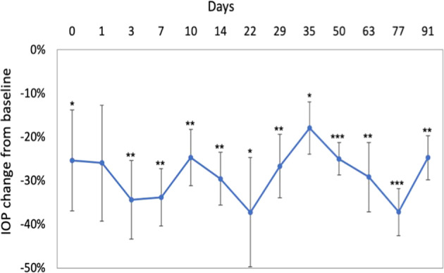
Results: VisiPlate demonstrated IOP-lowering of 20-40% compared to baseline at each time point over the course of 3 months in the nine implanted eyes. All eyes developed blebs over the implant, and fluorescein testing demonstrated fluid patency at 22 days post-implantation. Slit lamp and clinical observations showed that VisiPlate was well tolerated, with low levels of conjunctival congestion, conjunctival swelling, aqueous flare, hyphema, and iris involvement from surgery that resolved over time. At sacrifice time points of 93 days and 180 days, the only notable observations were mild levels of conjunctival congestion in implanted eyes. Histopathology showed minimal tissue response and no obvious inflammation, fibrosis, or necrosis around the implant.

Conclusions: The results of this in vivo study demonstrate the biocompatibility and IOP-lowering effect of a multichannel, ultrathin subconjunctival shunt in a rabbit model. The data suggest that VisiPlate may safely enhance aqueous outflow and significantly reduce intraocular pressure.


Conflict of interest statement

BWK: commercial relationship with Avisi Technologies, Co-Founder Avisi Technologies. EM: N/A. Thomas Barbolt: Commercial Relationship with Avisi Technologies, Consultant, Advisory Board. RAL: commercial relationship with Avisi Technologies, Advisory Board . IIA: commercial relationship with Avisi Technologies, Advisory Board. VL: commercial relationship with Avisi Technologies, Advisory Board. SMN: commercial relationship with Avisi Technologies, Consultant, Advisory Board. GG: commercial relationship with Avisi Technologies, VP Research and Development. EGM-E: Commercial Relationship with Avisi Technologies, Consultant, Advisory Board

Figures

Fig. 1
Fig. 1
Placement and structure of VisiPlate. A Anatomic placement of VisiPlate into the subconjunctival space. B Structure and dimensions of VisiPlate in millimeters, with scanning electron micrograph of VisiPlate’s surface topography of hexagonal corrugation and networked channels
Fig. 2
Fig. 2
Slit lamp exam photos from day 180 from Group 2 with implanted VisiPlate in the right eye (OD) treated with MMC and the left eye (OS) without MMC. Minimal inflammation was visible with appropriate placement of VisiPlate maintained in the subconjunctival space
Fig. 3
Fig. 3
Intraocular pressure measurements showing VisiPlate-implanted eyes with and without MMC injection in Group 2 (n = 3), presented as mean ± SE. After device implantation, IOP dropped to around 50% of baseline IOP post-operatively (around −6 to −7 mmHg, or 40–50% decrease from baseline) and remained low for the non-MMC-treated groups. MMC-treated eyes initially exhibited slightly greater IOP reductions (up to −8 mmHg or nearly 60% decrease from baseline) compared to eyes not treated with MMC; however, by month 6, MMC-treated eyes had returned to baseline. There was no significant difference between the MMC and non-MMC groups at any time point. IOPs were significantly lower (paired one-tailed Student’s t test, p < 0.05) than baseline at days 1, 3, 7, 10, 13, 30, 49, and 63 for the MMC + VisiPlate group, and at days 3, 7, 10, 14, 22, 30, 49, 63, 77, 91, 150 for the VisiPlate only group
Fig. 4
Fig. 4
Intraocular pressure decrease from baseline of eyes implanted with VisiPlate alone across all groups (n = 6), presented as mean ± SE. There was a significant reduction from baseline rabbit IOP of 15 mmHg exceeding the FDA’s efficacy threshold of 20% IOP reduction from baseline. *p < 0.05, **p < 0.01, ***p < 0.001, paired one-tailed Student’s t test
Fig. 5
Fig. 5
VisiPlate histology. Placement of VisiPlate in the subconjunctival space showing minimal tissue response at 180 days OD (A) and OS (B). The implant was present, located within a conjunctival pocket and extending into the anterior chamber, close to, but not adhered to, the corneal endothelium. No inflammation or fibrosis was associated with the portion of the implant present in the anterior chamber. Within the pocket, the implant folded once upon itself in one small area. Chronic inflammation (+1), consisting of very small numbers of macrophages and foreign body giant cells, were associated with the portion of the implant present within the pocket, particularly where the implant folded upon itself. Fibrosis and necrosis were not seen. Plastic-embedded sections from vicinity of implant location showing minimal tissue response (C). Higher magnification H&E view of implanted eye with giant cells on the surface of the implant (D). The implant seen within the anterior pocket at the bottom left of the image and extending into a conjunctival pocket extended up toward the top right with no obvious inflammation around the implant. Arrows show location of VisiPlate

References

    1. Parihar JK. Glaucoma: The 'Black hole' of irreversible blindness. Med J ArmedForces India. 2016;72:3–4. 10.1016/j.mjafi.2015.12.001. - PMC - PubMed
    1. Tham Y-C, Li X, Wong TY, Quigley HA, Aung T, Cheng C-Y. Global prevalence of glaucoma and projections of glaucoma burden through 2040: a systematic review and meta-analysis. Ophthalmology. 2014;121:2081–90. 10.1016/j.ophtha.2014.05.013. - PubMed
    1. Weinreb RN, Khaw PT. Primary open-angle glaucoma. Lancet. 2004;363:1711. doi: 10.1016/S0140-6736(04)16257-0. - DOI - PubMed
    1. Gedde SJ, et al. “Treatment outcomes in the Tube Versus Trabeculectomy (TVT) study after five years of follow-up.”. Am J Ophthalmol. 2012;153:789–803. doi: 10.1016/j.ajo.2011.10.026. - DOI - PMC - PubMed
    1. Goulet RJ, III, Phan AD, Cantor LB, WuDunn D. Efficacy of the Ahmed S2 Glaucoma valve compared with the Baerveldt 250 mm2 glaucoma implant. Ophthalmology. 2008;115:1141–7. doi: 10.1016/j.ophtha.2007.10.034. - DOI - PubMed

Substances