Review
. 2021 Nov 24;12(1):174.
doi: 10.1186/s13244-021-01118-y.

Communicating imaging findings in peritoneal mesothelioma: the impact of 'PAUSE' on surgical decision-making

Affiliations
Review

Communicating imaging findings in peritoneal mesothelioma: the impact of 'PAUSE' on surgical decision-making

Anuradha Chandramohan et al. Insights Imaging. .

Abstract

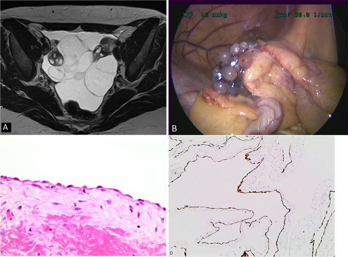
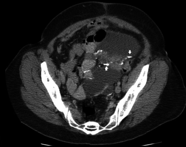
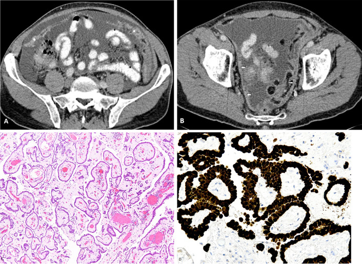
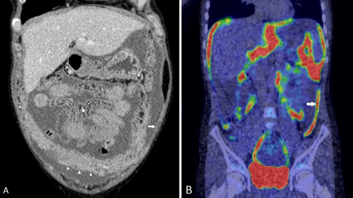
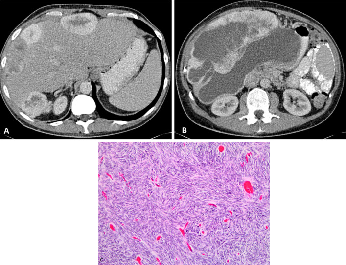
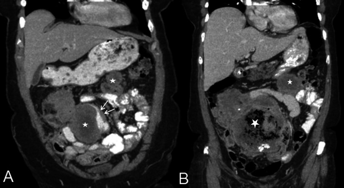
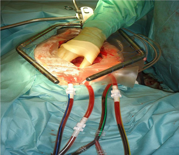
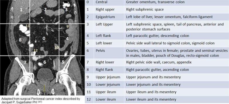
The peritoneal cavity is the second commonest site of mesothelioma after the pleural cavity. There are five histological types of peritoneal mesothelioma with variable symptomatology, clinical presentation and prognosis. Cystic mesothelioma is a borderline malignant neoplasm with a favourable prognosis, well-differentiated papillary mesothelioma is generally a low-grade malignancy, and all other varieties such as epithelioid, sarcomatoid and biphasic mesothelioma are highly malignant types of peritoneal mesothelioma with poor prognosis. Malignant peritoneal mesothelioma was considered inevitably fatal prior to the introduction of cytoreductive surgery (CRS) and hyperthermic intraperitoneal chemotherapy (HIPEC) in selected cases where long-term survival and cure could be achieved. However, the survival benefits following CRS and HIPEC mainly depend on completeness of cytoreduction, which come at the cost of high morbidity and potential mortality. Using the acronym 'PAUSE', we aimed at describing the key imaging findings that impact surgical decision-making in patients with peritoneal mesothelioma. PAUSE stands for peritoneal cancer index, ascites and abdominal wall disease, unfavourable sites of involvement, small bowel and mesenteric disease and extraperitoneal disease. Reporting components of 'PAUSE' is crucial for patient selection. Despite limitations of CT in accurately depicting the volume of disease, describing findings in terms of PAUSE plays an important role in excluding patients who might not benefit from CRS and HIPEC.

Keywords: Complete cytoreduction; Imaging; PAUSE; Peritoneal mesothelioma; Radiological peritoneal cancer index.


Conflict of interest statement

The authors have no financial or proprietary interests in any material discussed in this article.

Figures

Fig. 1
Fig. 1
ad 40-year-old female with cystic mesothelioma. a MRI T2 weighted axial image of the pelvis shows fluid intensity loculated cystic lesions surrounding the ovaries. b Intraoperative photograph showing the same findings. c High-power photomicrograph (H&E × 40) shows a flat layer of mesothelial cells lying on a loose fibrous stroma. d Low power photomicrograph (× 10) with calretinin immunohistochemical staining shows calretinin expression by the neoplastic cells which outline the shape of the cysts
Fig. 2
Fig. 2
ac 42-year-old male with diffuse form of cystic mesothelioma. a MRI T2 weighted axial image shows diffuse multi-cystic lesions in the peritoneal cavity. b CT through the pelvis shows fluid attenuation cystic lesions with fine non-enhancing septations. c Intra-operative photograph shows diffuse multi-loculated cystic masses in the diffusely filling the peritoneal cavity
Fig. 3
Fig. 3
CT axial image of a 47-year-old male patient with cystic mesothelioma showing nodular calcifications along the cyst walls (arrows)
Fig. 4
Fig. 4
ad 42-year-old female with well-differentiated papillary mesothelioma of the peritoneum. a, b CT axial images showing moderate volume ascites, plaque-like peritoneal thickening (arrow) and omental cake (*). c Low power photomicrograph (H&E, × 4) shows fibrovascular cores covered by a layer of cuboidal mesothelial cells. d High-power photomicrograph (calretinin immunostaining, × 20) showing expression of calretinin by the cells confirming mesothelial origin
Fig. 5
Fig. 5
ac 48-year-old male with ‘dry’ appearance of epithelioid type of malignant peritoneal mesothelioma. a, b Axial PET-CT images through the pelvis showing omental caking (*), nodular mesenteric fold thickening (arrows). b Axial image through the pelvis showing soft tissue density, FDG-avid, plaque-like, nodular soft tissue thickening of the pelvic peritoneum (*). c MRI T2 axial image showing T2 intermediate signal intensity, nodular soft tissue (*) along the pelvic peritoneum
Fig. 6
Fig. 6
ac 56-year-old male with ‘wet’ appearance of epithelioid peritoneal mesothelioma. a CT axial images showing omental cake (*) and ascites b Intraoperative photograph showing omental cake, peritoneal thickening and ascites. c High-power photomicrograph (H&E, × 20) demonstrates pleomorphic sheets of epithelioid cells with large nuclei, prominent nucleoli and eosinophilic cytoplasm. A few psammoma bodies are visible; one is marked with an arrowhead
Fig. 7
Fig. 7
a, b 47-year-old female with epithelioid type of malignant peritoneal mesothelioma. a Coronal CT image demonstrates diffuse plaque-like peritoneal thickening (white arrow), omental caking (arrow heads), ascites and tethered mesentery (*). b Coronal PET-CT shows FDG-avid plaque-like thickening (arrow) of the peritoneum
Fig. 8
Fig. 8
ac 57-year-old male with sarcomatoid peritoneal mesothelioma. a, b CT showing multiple intensely enhancing perihepatic peritoneal-based solid masses with central necrosis. c High-power photomicrograph (H&E × 20) showing spindle shaped tumour cells seen in sarcomatoid mesothelioma
Fig. 9
Fig. 9
a, b 78-year-old male with biphasic peritoneal mesothelioma. Serial CT scans show rapidly progressive disease. a Coronal CT showing large soft tissue mass in the mesentery (*) indenting and distorting the small bowel and causing eccentric bowel wall thickening (arrows). b Coronal CT section through the same site three months later shows increase in the size of the mass and air pockets within suggestive of contained bowel fistulation and disease progression
Fig. 10
Fig. 10
Photograph of hyperthermic intraperitoneal chemotherapy (HIPEC) procedure
Fig. 11
Fig. 11
Radiological peritoneal cancer index (rPCI) adapted from surgical peritoneal cancer index (PCI) described by Jacquet et al. [47]. This figure is being reused from author’s prior work published in clinical radiology [44] with permission from Elsevier
Fig. 12
Fig. 12
a, b: Abdominal wall disease in two different patients with malignant peritoneal mesothelioma. a CT of a patient with sarcomatoid type of peritoneal mesothelioma showing enhancing soft tissue nodule (*) in the abdominal wall. b CT of a patient with epithelioid mesothelioma shows a large lateral abdominal wall mass (*) abutting and tethering small bowel loop
Fig. 13
Fig. 13
a, b Images of two patients with epithelioid malignant peritoneal mesothelioma showing disease in the epigastric region. a Coronal CT with extensive peri-gastric disease (*) seen as soft tissue masses around the stomach. Also note the tethered mesentery. b Axial PET-CT images showing FDG-avid disease in the lesser omentum, lesser sac, porta (*) and peri-gastric region (arrows)
Fig. 14
Fig. 14
a, b: a PET-CT and (b) CT images of a patient with epithelioid malignant peritoneal mesothelioma showing nodular mesenteric fold thickening (white circle) and segmental small bowel obstruction(*)
Fig. 15
Fig. 15
CT scan, coronal image of a patient with advanced epithelioid malignant peritoneal mesothelioma showing a rind of soft tissue around the small bowel due to diffuse thickening of the small bowel serosa or the visceral peritoneum
Fig. 16
Fig. 16
ad CT images of different patients with malignant peritoneal mesothelioma showing findings of advanced mesenteric disease. a Nodular mesenteric fold thickening (arrows). b Sagittal CT showing soft tissue masses in the root of mesentery (*). c Mesenteric soft tissue mass (*) encasing small bowel. d Stellate mesentery on CT, which represents gross nodular thickening and retraction of the mesentery (*)
Fig. 17
Fig. 17
67-year-old male with biphasic type of malignant peritoneal mesothelioma. a, b CT coronal and sagittal reformatted images showing right perinephric soft tissue density mass (*), right hydronephrosis and ascites. This patient also had concurrent right intra-thoracic disease
Fig. 18
Fig. 18
a, b MRI images of a patient with biphasic malignant peritoneal mesothelioma. a Coronal MRI of the pelvis shows a large mixed solid cystic mass in the pelvis involving the left pelvic side wall (*). b Sagittal MRI of the pelvis shows the mass extending into presacral space (*). These constitute U2 findings which render complete cytoreduction unlikely
Fig. 19
Fig. 19
a, b Patient with epithelioid peritoneal mesothelioma. a CT shows ascites and subtle mesenteric nodularity (arrows). b Photograph from diagnostic laparoscopy showing diffuse mesenteric nodules (asterisk) and diffuse small bowel serosal nodules (black rectangle)
Fig. 20
Fig. 20
a, b Two different patients with biphasic malignant peritoneal mesothelioma. MRI shows (a) retroperitoneal (*) nodes and (b) left pelvic side wall lymph node (*). Note the disease extension into the left sciatic notch (b, arrow heads)
Fig. 21
Fig. 21
CT image of a patient with malignant peritoneal mesothelioma and concurrent pleural disease shows pleural based nodules (arrow) and pleural effusion

References

    1. Boffetta P. Epidemiology of peritoneal mesothelioma: a review. Ann Oncol. 2007;18:985–990. doi: 10.1093/annonc/mdl345. - DOI - PubMed
    1. Salo SA, Ilonen I, Laaksonen S, Myllärniemi M, Salo JA, Rantanen T. Epidemiology of malignant peritoneal mesothelioma: a population-based study. Cancer Epidemiol. 2017;51:81–86. doi: 10.1016/j.canep.2017.10.008. - DOI - PubMed
    1. Antman KH, Pomfret EA, Aisner J, MacIntyre J, Osteen RT, Greenberger JS. Peritoneal mesothelioma: natural history and response to chemotherapy. J Clin Oncol. 1983;1:386–391. doi: 10.1200/JCO.1983.1.6.386. - DOI - PubMed
    1. Brigand C, Monneuse O, Mohamed F, et al. Peritoneal mesothelioma treated by cytoreductive surgery and intraperitoneal hyperthermic chemotherapy: results of a prospective study. Ann Surg Oncol. 2006;13:405–412. doi: 10.1245/ASO.2006.05.041. - DOI - PubMed
    1. Spirtas R, Heineman EF, Bernstein L, et al. Malignant mesothelioma: attributable risk of asbestos exposure. Occup Environ Med. 1994;51:804–811. doi: 10.1136/oem.51.12.804. - DOI - PMC - PubMed

LinkOut - more resources