. 2021 Nov;10(13):e12164.
doi: 10.1002/jev2.12164.

Proteomic dissection of large extracellular vesicle surfaceome unravels interactive surface platform

Affiliations

Proteomic dissection of large extracellular vesicle surfaceome unravels interactive surface platform

Alin Rai et al. J Extracell Vesicles. 2021 Nov.

Abstract

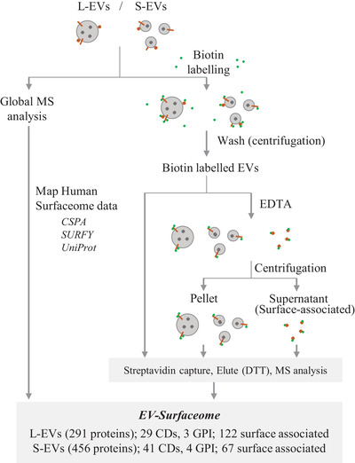
The extracellular vesicle (EV) surface proteome (surfaceome) acts as a fundamental signalling gateway by bridging intra- and extracellular signalling networks, dictates EVs' capacity to communicate and interact with their environment, and is a source of potential disease biomarkers and therapeutic targets. However, our understanding of surface protein composition of large EVs (L-EVs, 100-800 nm, mean 310 nm, ATP5F1A, ATP5F1B, DHX9, GOT2, HSPA5, HSPD1, MDH2, STOML2), a major EV-subtype that are distinct from small EVs (S-EVs, 30-150 nm, mean 110 nm, CD44, CD63, CD81, CD82, CD9, PDCD6IP, SDCBP, TSG101) remains limited. Using a membrane impermeant derivative of biotin to capture surface proteins coupled to mass spectrometry analysis, we show that out of 4143 proteins identified in density-gradient purified L-EVs (1.07-1.11 g/mL, from multiple cancer cell lines), 961 proteins are surface accessible. The surface molecular diversity of L-EVs include (i) bona fide plasma membrane anchored proteins (cluster of differentiation, transporters, receptors and GPI anchored proteins implicated in cell-cell and cell-ECM interactions); and (ii) membrane surface-associated proteins (that are released by divalent ion chelator EDTA) implicated in actin cytoskeleton regulation, junction organization, glycolysis and platelet activation. Ligand-receptor analysis of L-EV surfaceome (e.g., ITGAV/ITGB1) uncovered interactome spanning 172 experimentally verified cognate binding partners (e.g., ANGPTL3, PLG, and VTN) with highest tissue enrichment for liver. Assessment of biotin inaccessible L-EV proteome revealed enrichment for proteins belonging to COPI/II-coated ER/Golgi-derived vesicles and mitochondria. Additionally, despite common surface proteins identified in L-EVs and S-EVs, our data reveals surfaceome heterogeneity between the two EV-subtype. Collectively, our study provides critical insights into diverse proteins operating at the interactive platform of L-EVs and molecular leads for future studies seeking to decipher L-EV heterogeneity and function.

Keywords: extracellular vesicles; mass spectrometry-based proteomics; surface proteins; surfaceome; vesicle heterogeneity.


Figures

FIGURE 1
FIGURE 1
Isolation and characterization of large and small EVs. (a) Workflow for isolation of L‐EVs and S‐EVs. (b) Cryo‐electron microscopic images of L‐EVs and S‐EVs isolated from SW620 cells. (c) Histogram represents diameter of L‐EVs and S‐EVs based on cryo‐EM images. Data presented as mean ± s.e.m (standard error of mean). (d) Volcano plot of protein abundance between L‐EVs and S‐EVs using mass spectrometry‐based proteomics; comparisons of log2 fold changes versus p‐values (Student's t‐test). (e) Heat map of selected proteins enriched in L‐EVs or S‐EVs from three indicated cell lines (p‐value <0.05). (f) EnrichmentMap of Gene Ontology (cellular components) processes overrepresented in L‐EVs or S‐EVs. Node size represents gene number
FIGURE 2
FIGURE 2
Workflow for capturing surface proteins of large and small EVs. The workflow used in this study to define large and small EV surfaceome is outlined in Figure 3. Experimentally verified cell‐surface proteins (CSPA) (Bausch‐Fluck et al., 2015) and cellular surfaceome predicted by SURFY (Bausch‐Fluck et al., 2018) were mapped onto global EV protein profiles. Surface proteins were captured using membrane‐impermeant Sulfo‐NHS‐SS‐Biotin and subsequently identified by mass spectrometry. We also define a pool of EV‐surface associated proteome by treating EVs with EDTA and assessing released proteins. CD; cluster of differentiation, GPI; Glycosylphosphatidylinositol
FIGURE 3
FIGURE 3
Large EV s contain classical cellular surfaceome. Venn diagram of total proteins identified in (a). L‐EVs or (b). S‐EVs versus surface proteins reported in CSPA (Bausch‐Fluck et al., 2015), SURFY (Bausch‐Fluck et al., 2018) or UniProt. (c) Voronoi tree maps for L‐EV and S‐EV proteomes generated on wlab.ethz.ch/surfaceome. Light colour indicates low expression; dark colour indicates strong expression. White genes are not expressed. (d) Different classes of SURFY (Bausch‐Fluck et al., 2018) predicted cellular surface proteins identified in each EV‐subtype. (e) Volcano plot of differentially abundant surfaceome proteins in L‐EVs and S‐EVs
FIGURE 4
FIGURE 4
Proteome p rofiling of biotin accessible and inaccessible large EV proteomes. (a) Venn diagram of biotin‐captured surface proteins in L‐EVs versus surface proteins reported in CSPA (Bausch‐Fluck et al., 2015), SURFY (Bausch‐Fluck et al., 2018) or UniProt. (b) Bar plot of relative abundance of number of indicated classes of proteins identified versus total number of proteins identified in global or biotin‐captured surface proteome. (c) Venn diagram of biotin captured L‐EV surface proteins and global L‐EV proteome. Inner circle in L‐EV proteome represents a subset of SURFY/CSPA proteins detected in L‐EV global proteome. (d) Enrichment Map of Gene Ontology (cellular components) terms overrepresented in L‐EV surface proteome versus biotin inaccessible L‐EV proteome. (e) KEGG and Reactome pathways overrepresented in L‐EV surface proteome versus biotin inaccessible L‐EV proteome
FIGURE 5
FIGURE 5
Surface‐associated proteins in large EVs. (a) Venn diagram of proteins identified in L‐EV surface proteome, and proteins that remain either bound or are released from L‐EV surface following EDTA treatment. Bar plot of protein numbers of indicated classes that are either released (b) or remain bound (c) with EVs following EDTA treatment. (d) L‐EV biotin surface proteome was divided into CSPA/SURFY proteins (i.e., classical cellular surfaceome) or non‐CSPA/SURFY proteins (i.e., non‐classical surface proteins); Venn diagram reveals pool of these proteins that remain either bound or are released from L‐EV surface following EDTA treatment. (e) EnrichmentMap of KEGG and Reactome pathways overrepresented in L‐EV proteins that remain either bound or are released from L‐EV surface following EDTA treatment. (f) GeneMania‐based radial interaction map of membrane‐bound and membrane‐associated (i.e., released) proteins in L‐EVs. Nodes represent the proteins, and the edges represent evidence‐based direct physical interactions. Pathway involvement for indicated proteins are asterisk‐colour coded
FIGURE 6
FIGURE 6
Receptor‐ligand interactome of large EV surface proteome. (a) Venn diagram of L‐EV biotin surface proteins (classical) and their experimentally‐verified cognate binding partners based on Cellinker that catalogues literature‐supported ligand‐receptor interactions (C. Zhang, Wang, et al., 2021). (b) Distribution of interaction‐type identified in L‐EVs. (c) Sankey diagram of ligand‐receptor interactions identified in L‐EVs; complete list provided in Table S7. (d) Bar plot represents tissue enrichment (based on DAVID analysis) of 172 cognate interacting partners of L‐EV surface proteins. (e) Cellinker‐based interacting ligands identified for L‐EV surface ITGB1/ITGAV proteins. (f) RNA expression and tissue specificity for ANGPTL3, PLG and VTN in different human tissues was obtained from The Human Protein Atlas (http://www.proteinatlas.org, image credit: Human Protein Atlas [Uhlén et al., 2015])

References

    1. Al‐Nedawi, K. , Meehan, B. , Micallef, J. , Lhotak, V. , May, L. , Guha, A. , & Rak, J. (2008). Intercellular transfer of the oncogenic receptor EGFRvIII by microvesicles derived from tumour cells. Nature Cell Biology, 10, 619–624. - PubMed
    1. Bausch‐Fluck, D. , Goldmann, U. , Müller, S. , Van Oostrum, M. , Müller, M. , Schubert, O. T. , & Wollscheid, B. (2018). The in silico human surfaceome. PNAS, 115, E10988–E10997. - PMC - PubMed
    1. Bausch‐Fluck, D. , Hofmann, A. , Bock, T. , Frei, A. P. , Cerciello, F. , Jacobs, A. , Moest, H. , Omasits, U. , Gundry, R. L. , Yoon, C. , Schiess, R. , Schmidt, A. , Mirkowska, P. , Härtlová, A. , Van Eyk, J. E. , Bourquin, J.‐P. , Aebersold, R. , Boheler, K. R. , Zandstra, P. , & Wollscheid, B. (2015). A mass spectrometric‐derived cell surface protein atlas. Plos One, 10, e0121314. - PMC - PubMed
    1. Bebawy, M. , Combes, V. , Lee, E. , Jaiswal, R. , Gong, J. , Bonhoure, A. , Grau, G. E. R. (2009). Membrane microparticles mediate transfer of P‐glycoprotein to drug sensitive cancer cells. Leukemia, 23, 1643–1649. - PubMed
    1. Bellin, G. , Gardin, C. , Ferroni, L. , Chachques, J. , Rogante, M. , Mitrečić, D. , Ferrari, R. , & Zavan, B. (2019). Exosome in cardiovascular diseases: A complex world full of hope. Cells, 8, 166. - PMC - PubMed

Publication types

MeSH terms