. 2021 Dec 8;143(48):20095-20108.
doi: 10.1021/jacs.1c06167. Epub 2021 Nov 24.

Site-Specific Labeling of Endogenous Proteins Using CoLDR Chemistry

Affiliations

Site-Specific Labeling of Endogenous Proteins Using CoLDR Chemistry

Rambabu N Reddi et al. J Am Chem Soc. .

Abstract

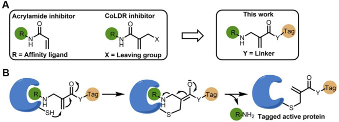
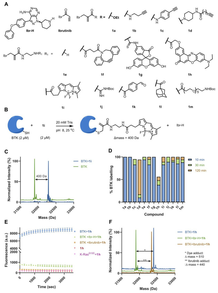
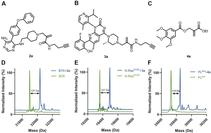
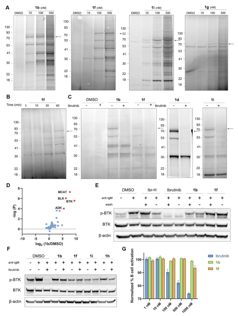
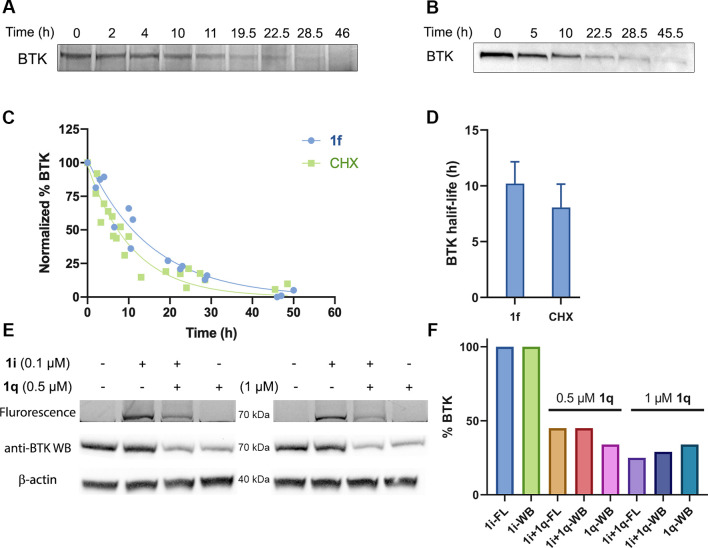
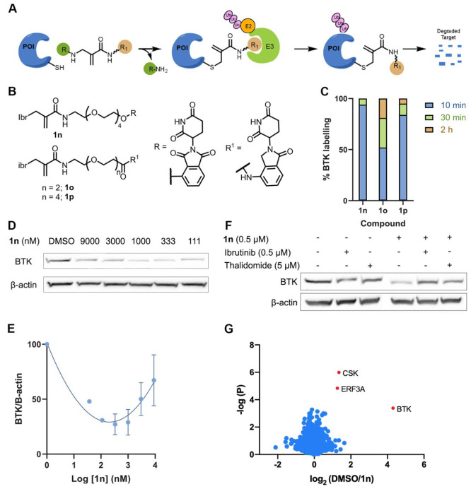
Chemical modifications of native proteins can affect their stability, activity, interactions, localization, and more. However, there are few nongenetic methods for the installation of chemical modifications at a specific protein site in cells. Here we report a covalent ligand directed release (CoLDR) site-specific labeling strategy, which enables the installation of a variety of functional tags on a target protein while releasing the directing ligand. Using this approach, we were able to label various proteins such as BTK, K-RasG12C, and SARS-CoV-2 PLpro with different tags. For BTK we have shown selective labeling in cells of both alkyne and fluorophores tags. Protein labeling by traditional affinity methods often inhibits protein activity since the directing ligand permanently occupies the target binding pocket. We have shown that using CoLDR chemistry, modification of BTK by these probes in cells preserves its activity. We demonstrated several applications for this approach including determining the half-life of BTK in its native environment with minimal perturbation, as well as quantification of BTK degradation by a noncovalent proteolysis targeting chimera (PROTAC) by in-gel fluorescence. Using an environment-sensitive "turn-on" fluorescent probe, we were able to monitor ligand binding to the active site of BTK. Finally, we have demonstrated efficient CoLDR-based BTK PROTACs (DC50 < 100 nM), which installed a CRBN binder onto BTK. This approach joins very few available labeling strategies that maintain the target protein activity and thus makes an important addition to the toolbox of chemical biology.


Conflict of interest statement

The authors declare the following competing financial interest(s): N.L., R.N.R., A.R., and E.R. are inventors on a patent application describing this technology.

Figures

Figure 1
Figure 1
Development of ligand-directed cysteine labeling probes. (A) By reversing the directionality of our previously developed CoLDR chemistry, we generate probes that place the electrophilic carbon in the exact same position but now release the protein recognition moiety (R; typically an inhibitor). (B) Schematic representation of the reaction of a target cysteine with a substituted α-methacrylamide through CoLDR chemistry.
Figure 2
Figure 2
Site-selective labeling of BTK using CoLDR chemistry. (A) Chemical structures of ibrutinib-directed methacrylamides with various functional tags. (B) Typical example of the reaction of BTK (2 μM) with 1i (2 μM) in a 20 mM Tris buffer at pH 8, 25 °C. (C) Deconvoluted LC/MS spectrum shows the labeling of a BODIPY probe and demonstrates Ibr-H leaving. (D) Percent of labeling of BTK (2 μM) with the probes (1a1m; 2 μM) at 10, 30, and 120 min in 20 mM Tris buffer at pH 8, 25 °C. (E) Kinetics of the increase in fluorescence intensity measured at Ex/Em = 550/620 nm (n = 4) upon addition of BTK (2 μM) to 1h (2 μM) in 20 mM Tris buffer at pH 8, 37 °C (blue). Control experiments without BTK (red), preincubation of ibrutinib (4 μM) and Ibr-H (4 μM) prior to adding 1h (green and orange, respectively), and incubation of K-RasG12C (pink) with 1h show no fluorescence. (F) Deconvoluted LC/MS spectra for BTK incubated with 1h at the end of the fluorescence measurement (shown in E). The adduct mass corresponds to a labeling event in which the Ibr-H moiety was released, validating the proposed mechanism.
Figure 3
Figure 3
Selective labeling of various target proteins. Structures of alkyne/ester labeling probes for (A) BTK, (B) K-RasG12C, and (C) SARS-CoV-2 PLpro. Deconvoluted LC/MS spectra for (D) BTK (2 μM) incubated with 2a (2 μM) in 20 mM Tris buffer at pH 8, 25 °C, 10 min, (E) K-RasG12C (10 μM) incubated with 3a (100 μM) in 20 mM Tris at pH 8, 37 °C, 16 h, and (F) PLpro (2 μM) incubated with 4a (10 μM) in 50 mM Tris at pH 8, 25 °C, 16 h. The adduct masses correspond to a labeling event in which the ligand was released.
Figure 4
Figure 4
Labeling BTK with CoLDR probes does not inhibit its activity in cells. (A) Cellular labeling profile of 1b, 1f, and 1i after 2 h of incubation with Mino cells and 1g in Mino cell lysate. 1b and 1f samples were further reacted with TAMRA-azide in lysate before imaging. An arrow indicates BTK’s MW. (B) Time-dependent labeling profile of 1f with BTK after incubation of Mino cells with 100 nM probe followed by a click reaction with TAMRA-azide in lysate prior to imaging. (C) Competition experiment of 1b, 1d, 1f, and 1i with ibrutinib. The cells were preincubated for 30 min with either 0.1% DMSO or 1 μM ibrutinib, followed by 2 h of incubation with 200 nM 1b or 1f or 100 nM 1d or 1i. (D) Mino cells were incubated with 0.1% DMSO or 1b (100 nM). Samples were further reacted with biotin-azide in lysate, followed by enrichment, trypsin digestion, and peptide identification by LC/MS/MS. The log(fold-ratio) of proteins enriched by 1b over DMSO is plotted as a function of statistical significance. BTK is clearly identified as the most enriched target; additional prominent targets that correspond to bands identified by in-gel fluorescence (panel C) are indicated. (E) BTK activity assay in Mino cells as measured by autophosphorylation of BTK. The cells were incubated for 1 h with either 0.1% DMSO, 1 μM ibrutinib, 1 μM Ibr-H, or 100 nM 1b, 1f, 1h, or 1i. The cells were either washed or not before induction of BTK activity by anti-IgM. (F) BTK activity assay: Mino cells were incubated for 2 h with either DMSO or 1 μM 1b, 1f, 1i, and 1h, washed, and then incubated for 45 min with ibrutinib (100 nM). The cells were washed again before induction of BTK activity by anti-IgM. The CoLDR probes were able to rescue BTK activity from inhibition by ibrutinib. (G) Primary B cell activation induced by anti-IgM after 24 h of treatment with increasing doses of either ibrutinib, 1b, or 1f, showing no inhibition of the CoLDR probes.
Figure 5
Figure 5
Measurement of BTK half-life. (A) Half-life measurement of BTK using 1f. Mino cells were pulse-labeled with 100 nM 1f for 1 h and were then washed to remove the excess probe. Cells were harvested at the indicated time points, and lysates were reacted with TAMRA-azide. The signal of BTK was quantified, and the half-life was calculated. (B) Half-life measurement of BTK with the cycloheximide (CHX) assay, using 20 μg/mL cycloheximide. (C) Quantification of BTK levels in A and B (by normalization to the protein concentration) in Mino cells (1f: n = 3, CHX: n = 4). (D) Calculated half-life by both methods, presented as mean ± SD. (E) Degradation of BTK labeled with 1i using PROTAC 1q. Mino cells were incubated with 1i (100 nM), then washed to remove the excess probe, again incubated with PROTAC 1q for 2 h at 0.5 and 1 μM, and then lysed. Samples are subjected to in-gel fluorescence (FL) and Western blot (WB). (F) Quantification of BTK levels in panel E (normalization to the β-actin has been done for Western blot).
Figure 6
Figure 6
Measurement of induced degradation by CoLDR PROTACs. (A) Schematic representation of target degradation using CoLDR PROTACS. (B) Structure of CoLDR-based BTK PROTACS. (C) In vitro labeling of BTK (2 μM) with 1n1p (2 μM) in 20 mM Tris buffer at pH 8, 25 °C. (D) Western blot evaluation of BTK levels in Mino cells in response to various concentrations of 1n after 24 h of incubation. (E) Quantification of BTK levels in D by normalization to the β-actin house-keeping gene in Mino cells. DC50 and Dmax were calculated by fitting the data to a second-order polynomial using the Prism software. (F) Mino cells were pretreated for 2 h with either ibrutinib/thalidomide-OH or DMSO before treatment with a BTK PROTAC for 24 h (n = 2). Subsequently, BTK levels were measured via Western blot. (G) Mino cells were treated for 24 h with either 0.1% DMSO or 1n (500 nM) in 4 replicates. Lysates were subjected to trypsin digestion and peptide identification by LC/MS/MS. The Log2(fold-ratio) of proteins enriched in the DMSO samples over 1n-treated samples is plotted as a function of statistical significance. Significantly degraded proteins are indicated in red and defined as Log2(DMSO/1n) > 1 and p-value < 0.01.

References

    1. Griffin B. A.; Adams S. R.; Tsien R. Y. Specific Covalent Labeling of Recombinant Protein Molecules inside Live Cells. Science 1998, 281 (5374), 269–272. 10.1126/science.281.5374.269. - DOI - PubMed
    1. Xue L.; Karpenko I. A.; Hiblot J.; Johnsson K. Imaging and Manipulating Proteins in Live Cells through Covalent Labeling. Nat. Chem. Biol. 2015, 11 (12), 917–923. 10.1038/nchembio.1959. - DOI - PubMed
    1. Nischan N.; Hackenberger C. P. R. Site-Specific PEGylation of Proteins: Recent Developments. J. Org. Chem. 2014, 79 (22), 10727–10733. 10.1021/jo502136n. - DOI - PubMed
    1. Tsai Y.-H.; Essig S.; James J. R.; Lang K.; Chin J. W. Selective, Rapid and Optically Switchable Regulation of Protein Function in Live Mammalian Cells. Nat. Chem. 2015, 7 (7), 554–561. 10.1038/nchem.2253. - DOI - PMC - PubMed
    1. Yang S.-T.; Lim S. I.; Kiessling V.; Kwon I.; Tamm L. K. Site-Specific Fluorescent Labeling to Visualize Membrane Translocation of a Myristoyl Switch Protein. Sci. Rep. 2016, 6, 32866.10.1038/srep32866. - DOI - PMC - PubMed

Publication types

MeSH terms