. 2022 Feb 8;88(3):e0170521.
doi: 10.1128/AEM.01705-21. Epub 2021 Nov 24.

Type IX Secretion System Effectors and Virulence of the Model Flavobacterium columnare Strain MS-FC-4

Affiliations

Type IX Secretion System Effectors and Virulence of the Model Flavobacterium columnare Strain MS-FC-4

Nicole C Thunes et al. Appl Environ Microbiol. .

Abstract

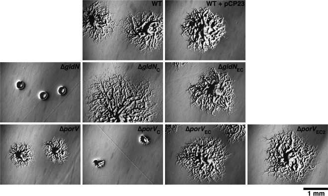
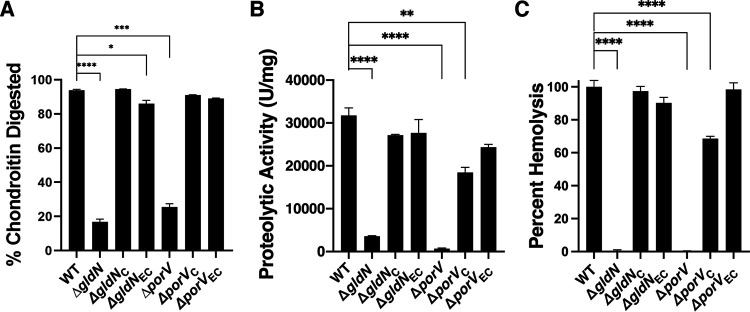
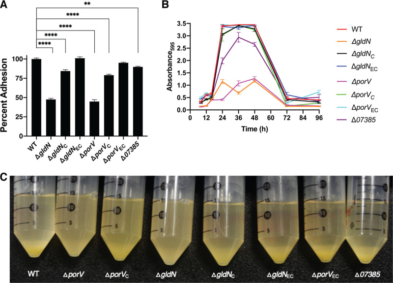
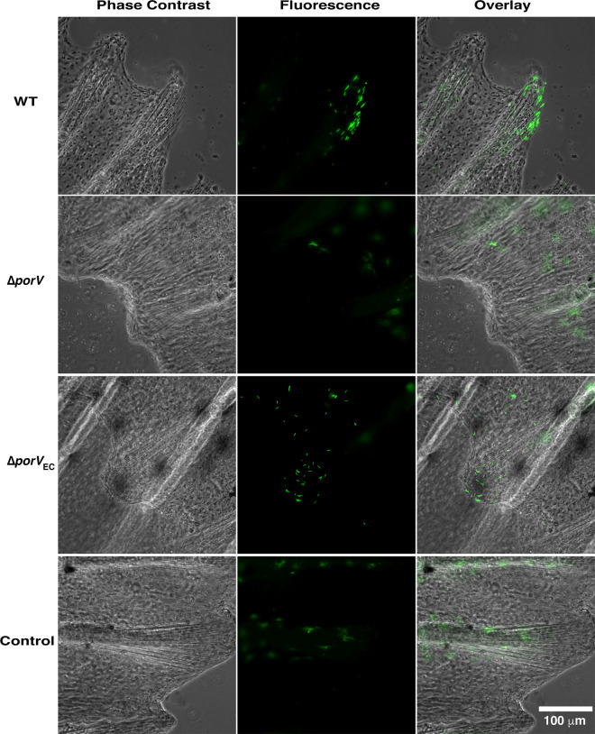
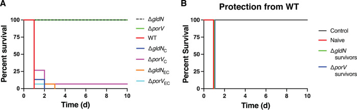
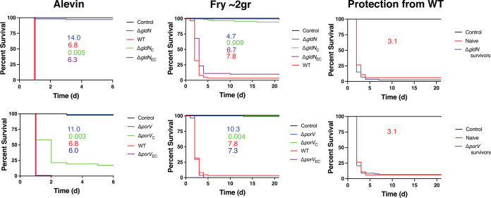
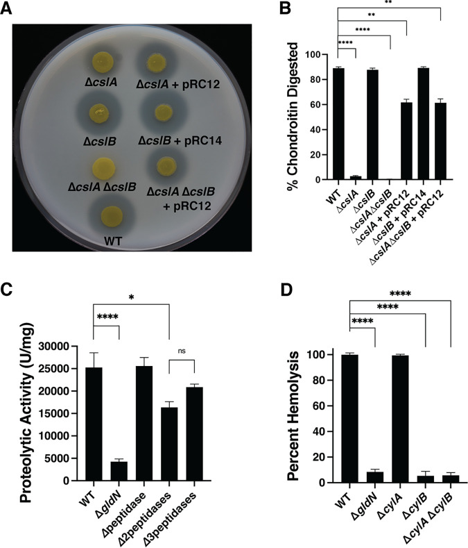
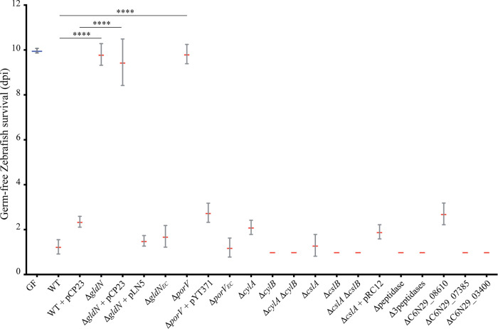
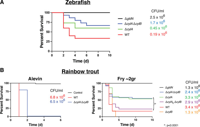
Flavobacterium columnare causes columnaris disease in wild and cultured freshwater fish and is a major problem for sustainable aquaculture worldwide. The F. columnare type IX secretion system (T9SS) secretes many proteins and is required for virulence. The T9SS component GldN is required for secretion and gliding motility over surfaces. Genetic manipulation of F. columnare is inefficient, which has impeded identification of secreted proteins that are critical for virulence. Here, we identified a virulent wild-type F. columnare strain (MS-FC-4) that is highly amenable to genetic manipulation. This facilitated isolation and characterization of two deletion mutants lacking core components of the T9SS. Deletion of gldN disrupted protein secretion and gliding motility and eliminated virulence in zebrafish and rainbow trout. Deletion of porV disrupted secretion and virulence but not motility. Both mutants exhibited decreased extracellular proteolytic, hemolytic, and chondroitin sulfate lyase activities. They also exhibited decreased biofilm formation and decreased attachment to fish fins and other surfaces. Using genomic and proteomic approaches, we identified proteins secreted by the T9SS. We deleted 10 genes encoding secreted proteins and characterized the virulence of mutants lacking individual or multiple secreted proteins. A mutant lacking two genes encoding predicted peptidases exhibited reduced virulence in rainbow trout, and mutants lacking a predicted cytolysin showed reduced virulence in zebrafish and rainbow trout. The results establish F. columnare strain MS-FC-4 as a genetically amenable model to identify virulence factors. This may aid development of measures to control columnaris disease and impact fish health and sustainable aquaculture. IMPORTANCE Flavobacterium columnare causes columnaris disease in wild and aquaculture-reared freshwater fish and is a major problem for aquaculture. Little is known regarding the virulence factors involved in this disease, and control measures are inadequate. The type IX secretion system (T9SS) secretes many proteins and is required for virulence, but the secreted virulence factors are not known. We identified a strain of F. columnare (MS-FC-4) that is well suited for genetic manipulation. The components of the T9SS and the proteins secreted by this system were identified. Deletion of core T9SS genes eliminated virulence. Genes encoding 10 secreted proteins were deleted. Deletion of two peptidase-encoding genes resulted in decreased virulence in rainbow trout, and deletion of a cytolysin-encoding gene resulted in decreased virulence in rainbow trout and zebrafish. Secreted peptidases and cytolysins are likely virulence factors and are targets for the development of control measures.

Keywords: columnaris disease; fish pathogen; flavobacterium; type IX secretion system.


Conflict of interest statement

The authors declare no conflict of interest.

Figures

FIG 1
FIG 1
Gliding of wild-type and mutant cells on glass. Cells were grown in TYES at 28°C to mid-exponential phase (OD600 approximately 0.5). Ten microliters of culture were introduced into a glass tunnel slide and observed for motility using an Olympus BH-2 phase-contrast microscope. Wild-type (WT) F. columnare, ΔgldN mutant, ΔporV mutant, ΔgldN complemented with wild-type gldN on pLN5 (ΔgldNC), and ΔporV complemented with wild-type porV on pYT371 (ΔporVC) are shown. This figure also includes ΔporV ectopically complemented with wild-type porV inserted at a neutral site on the chromosome and expressed from the ompA promoter (ΔporVEC). In each case, a series of images were taken using a Photometrics CoolSNAPcf2 camera. Individual frames were colored from red (time zero) to yellow, green, cyan, and finally blue (40 s) and integrated into one image, resulting in “rainbow traces” of gliding cells. The top six panels (in grayscale) show the first frame for each strain. The bottom six panels show the corresponding 40-s rainbow traces. White cells in the bottom panel correspond to cells that exhibited little, if any, net movement. Bar at lower right (20 μm) applies to all panels. The rainbow traces correspond to the sequences in Movie S1 and Movie S2 in the supplemental material.
FIG 2
FIG 2
Colonies of wild-type, mutant, and complemented strains of F. columnare. Colonies of wild type (WT), ΔgldN mutant, ΔgldN mutant complemented with wild-type gldN on plasmid pLN5 (ΔgldNC), ΔporV mutant, and ΔporV mutant complemented with wild-type porV on plasmid pYT371 (ΔporVC). This figure also includes colonies for ΔgldN ectopically complemented with wild-type gldN at a neutral site on the chromosome and expressed from the ompA promoter (ΔgldNEC), ΔporV complemented with wild-type porV at a neutral site on the chromosome and expressed from the ompA promoter (ΔporVEC), and ΔporV complemented with wild-type porV at a neutral site on the chromosome and expressed from the native porV promoter (ΔporVEC2). Colonies arose from cells incubated for 45 h at 30°C on one-quarter strength TYES agar. Photomicrographs were taken with a Photometrics CoolSNAPcf2 camera mounted on an Olympus IMT-2 phase-contrast microscope. Bar (1 mm) applies to all images.
FIG 3
FIG 3
Chondroitin sulfate lyase, proteolysis, and hemolytic activities of material secreted by wild type (WT), ΔgldN mutant, ΔporV mutant, and complemented mutants. The gldN mutant was complemented by pLN5 (ΔgldNC) or by insertion of gldN on the chromosome (ΔgldNEC). The porV mutant was complemented by pYT371 (ΔporVC) or by insertion of porV on the chromosome (ΔporVEC). (A) Secreted chondroitin sulfate lyase activity. (B) Secreted proteolytic activity. (C) Secreted hemolytic activity normalized to wild type. Statistics correspond to one-way ANOVA with Tukey posttest comparing all conditions to wild type. ****, P < 0.0001; ***, P < 0.001; **, P < 0.01; *, P < 0.05; absence of star, nonsignificant.
FIG 4
FIG 4
Adhesion, biofilm, and sedimentation of wild-type, mutant, and complemented strains. Strains examined were wild-type F. columnare MS-FC-4 (WT), ΔgldN mutant, ΔporV mutant, ΔgldN mutant complemented with pLN5 (ΔgldNC), ΔgldN complemented with wild-type gldN on the chromosome (ΔgldNEC), ΔporV mutant complemented with pYT371 (ΔporVC), ΔporV complemented with wild-type porV on the chromosome (ΔporVEC), and ΔC6N29_07385. (A) Adhesion to polystyrene after 3 h of incubation at 30°C without shaking as determined by staining with crystal violet and measuring absorbance at 595 nm. Adhesion shown in relation to the wild-type strain, which was set as 100. (B) Biofilm formation on polystyrene of cells in half-strength TYES broth incubated for 96 h at 30°C without shaking. (C) Cell sedimentation. Cells grown in half-strength TYES broth for 24 h at 28°C with shaking at 200 rpm were transferred to a centrifuge tube and allowed to stand at room temperature for 5 h without shaking before being photographed. Statistics correspond to one-way ANOVA with Tukey posttest comparing all conditions to wild type. ****, P < 0.0001; **, P < 0.01; absence of star, nonsignificant.
FIG 5
FIG 5
Adhesion to zebrafish fins of wild type, ΔporV mutant, and ΔporV mutant complemented with porV inserted ectopically on the chromosome and expressed from the ompA promoter (ΔporVEC). Cells of each strain carried pNT67, which expresses GFP. Live fish were exposed to the F. columnare strains for 60 min and rinsed twice in clean water, and pectoral fins were examined for attached cells by fluorescence microscopy. The bottom row shows part of a fin from a fish that was not exposed to F. columnare cells and illustrates the background fluorescence not associated with F. columnare cells. (Left column) Phase contrast images. (Middle column) Fluorescence images. (Right column) Composite images. Six fins were examined for each strain. Representative images are shown. Bar indicating 100 μm applies to all images.
FIG 6
FIG 6
T9SS is required for virulence in zebrafish. Zebrafish were exposed by immersion to F. columnare strains for 1 h at 28°C and transferred to fresh water, and percent survival was monitored for 10 days. (A) Strains examined were wild-type F. columnare MS-FC-4 (WT), ΔgldN mutant, ΔporV mutant, ΔgldN mutant complemented with plasmid pLN5 (ΔgldNC), ΔgldN ectopically complemented with wild-type gldN at a neutral site on the chromosome and expressed from the ompA promoter (ΔgldNEC), ΔporV mutant complemented with plasmid pYT371 (ΔporVC), and ΔporV complemented with wild-type porV at a neutral site on the chromosome and expressed from the ompA promoter (ΔporVEC). The final challenge concentrations were 2.3 × 106 CFU/mL (WT), 2.8 × 106 CFU/mL (ΔgldN), 4.2 × 106 CFU/mL (ΔporV), 1.9 × 106 CFU/mL (ΔgldNC), 2.5 × 106 CFU/mL (ΔgldNEC), 1.9 × 105 CFU/mL (ΔporVC), and 1.4 × 106 CFU/mL (ΔporVEC). Fifteen fish were challenged with each strain as indicated in Materials and Methods. Five control fish that were exposed to an equal amount of growth medium without F. columnare cells were also included, and each of these control fish survived. (B) Fish that survived exposure to ΔgldNgldN survivors) and ΔporVporV survivors) were examined for resistance to wild-type cells. Naive fish, ΔgldN survivors, and ΔporV survivors were maintained for 28 days and then exposed to wild-type cells at 2.9 × 106 CFU/mL for 1 h. Survival was monitored for 10 days. “Control” indicates three additional fish that had survived exposure to ΔgldN and three fish that had survived exposure to ΔporV that were exposed to an equivalent volume of TYES medium instead of to wild-type cells.
FIG 7
FIG 7
T9SS is required for virulence in rainbow trout. Rainbow trout were exposed by immersion to F. columnare strains, and percent survival was monitored for 21 days. Strains examined were wild-type F. columnare MS-FC-4 (WT), ΔgldN mutant, ΔporV mutant, ΔgldN mutant complemented with plasmid pLN5 (ΔgldNC), ΔgldN ectopically complemented with wild-type gldN at a neutral site on the chromosome and expressed from the ompA promoter (ΔgldNEC), ΔporV mutant complemented with plasmid pYT371 (ΔporVC), and ΔporV complemented with wild-type porV at a neutral site on the chromosome and expressed from the ompA promoter (ΔporVEC). The final challenge concentrations for each strain are given in the corresponding color and indicate CFU × 106/mL (for example, 6.8 × 106 CFU/mL for wild-type challenges of alevin). Results for WT are in red, TYES control in black, ΔporV or ΔgldN in blue, plasmid complementation in green, and ectopic integrated complemention in purple. (Left column) Rainbow trout alevin. (Middle column) Rainbow trout fry. (Right column) Naive fry and fry that survived exposure to ΔgldNgldN survivors) and ΔporVporV survivors) were examined for later resistance to challenge with wild-type cells.
FIG 8
FIG 8
Chondroitin sulfate lyase, proteolysis, and hemolytic activities of material secreted by wild-type and mutant strains. (A) Qualitative plate assay for chondroitin sulfate lyase activities of wild type, mutants (ΔcslA, ΔcslB, and ΔcslA ΔcslB), and mutants complemented with pRC12, which carries cslA, or with pRC14, which carries cslB. (B) Quantitative assay for chondroitin sulfate lyase activities for the same strains as in panel A as determined by measuring the amount of undigested chondroitin sulfate A in each well of a 96-well plate after incubating the cells for 30 min at 30°C. Measurements were taken in triplicate. (C) Secreted proteolytic activities of wild type, ΔgldN, ΔC6N29_05800 (Δpeptidase), ΔC6N29_11545-C6N29_11550 (Δ2peptidases), and ΔC6N29_05800 ΔC6N29_11545-C6N29_11550 (Δ3peptidases). (D) Hemolysis activities of wild type and mutants (ΔcylA, ΔcylB, and ΔcylA ΔcylB). Statistics correspond to one-way ANOVA with Tukey posttest comparing all conditions to wild type. ****, P < 0.0001; ***, P < 0.001; **, P < 0.01; *, P < 0.05; absence of star, nonsignificant.
FIG 9
FIG 9
Challenge of germfree zebrafish larvae with F. columnare strains. Survival of germfree zebrafish larvae exposed to wild-type, mutant, or complemented F. columnare strains. All strains were derived from wild-type (WT) F. columnare strain MS-FC-4. “Δpeptidase” indicates deletion of C6N29_05800, and “Δ3peptidase” indicates deletion of C6N29_05800, C6N29_11545, and C6N29_11550. Zebrafish larvae were infected at 6 days postfertilization by immersion for 3 h with 5 × 104 CFU/mL. Zero days postinfection (dpi) corresponds to the day of infection. Mean survival is represented by a thick horizontal bar with standard deviation. For each condition, n = 10 to 12 zebrafish larvae. Larval mortality rate was monitored daily, and surviving fish were euthanized at day 9 postinfection. Statistics correspond to unpaired, nonparametric Mann-Whitney test comparing all conditions to noninfected larvae (GF). ****, P < 0.0001; ***, P < 0.001; absence of star, nonsignificant. Blue mean bar corresponds to larvae not exposed to the pathogen (GF), and red mean bars correspond to exposed larvae.
FIG 10
FIG 10
Deletion of two genes encoding predicted peptidases resulted in partial defect in virulence in rainbow trout. Rainbow trout were exposed by immersion to F. columnare strains, and percent survival was monitored. Strains examined were wild-type F. columnare MS-FC-4 (WT, red), ΔgldN (black), ΔC6N29_05800 (Δpeptidase, purple), ΔC6N29_11545-C6N29_11550 (Δ2peptidases, green), and ΔC6N29_05800 ΔC6N29_11545-C6N29_11550 (Δ3peptidases, blue). The final challenge concentrations for each strain are given in the corresponding color. (A) Challenge of rainbow trout alevin. (B) Challenge of rainbow trout fry. “Control” indicates fish exposed to equivalent amount of TYES growth medium instead of to F. columnare.
FIG 11
FIG 11
Role of the predicted cytolysins CylA and CylB in virulence. Fish were exposed by immersion to F. columnare strains, and percent survival was monitored. Strains examined were wild-type F. columnare MS-FC-4 (WT, red), ΔgldN (black), ΔcylA (green), ΔcylB (orange), ΔcylA ΔcylB double mutant (blue), and ΔcylA ΔcylB complemented with pNT69, which carries cylAcylAC ΔcylB, purple). The final challenge concentrations for each strain are given in the corresponding color. (A) Challenge of zebrafish. Fifteen zebrafish were challenged with each strain. (B) Challenge of rainbow trout alevin (left) and fry (right). “Control” indicates fish exposed to equivalent amount of TYES growth medium instead of to F. columnare.

Similar articles

Cited by

References

    1. Davis HS. 1922. A new bacterial disease of freshwater fishes. Bull US Bureau Fish 38:261–280.
    1. Declercq AM, Haesebrouck F, Van den Broeck W, Bossier P, Decostere A. 2013. Columnaris disease in fish: a review with emphasis on bacterium-host interactions. Vet Res 44:27. 10.1186/1297-9716-44-27. - DOI - PMC - PubMed
    1. Bullock GL, Hsu TC, Shotts EB. 1986. Columnaris disease of fishes. Fish Disease Leaflet 72. U.S. Fish and Wildlife Service, U.S. Department of the Interior, Washington, DC.
    1. Wagner BA, Wise DJ, Khoo LH, Terhune JS. 2002. The epidemiology of bacterial diseases in food-size channel catfish. J Aquat Anim Health 14:263–272. 10.1577/1548-8667(2002)014<0263:TEOBDI>2.0.CO;2. - DOI - PubMed
    1. LaFrentz BR, Waldbieser GC, Welch TJ, Shoemaker CA. 2014. Intragenomic heterogeneity in the 16S rRNA genes of Flavobacterium columnare and standard protocol for genomovar assignment. J Fish Dis 37:657–669. 10.1111/jfd.12166. - DOI - PubMed

Publication types

Supplementary concepts

LinkOut - more resources