Expression of Spred2 in the urothelial tumorigenesis of the urinary bladder
- PMID: 34818323
- PMCID: PMC8612556
- DOI: 10.1371/journal.pone.0254289
Expression of Spred2 in the urothelial tumorigenesis of the urinary bladder
Abstract
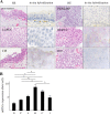
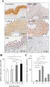
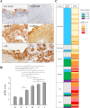
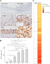
Aberrant activation of the Ras/Raf/ERK (extracellular-signal-regulated kinase)-MAPK (mitogen-activated protein kinase) pathway is involved in the progression of cancer, including urothelial carcinoma; but the negative regulation remains unclear. In the present study, we investigated pathological expression of Spred2 (Sprouty-related EVH1 domain-containing protein 2), a negative regulator of the Ras/Raf/ERK-MAPK pathway, and the relation to ERK activation and Ki67 index in various categories of 275 urothelial tumors obtained from clinical patients. In situ hybridization demonstrated that Spred2 mRNA was highly expressed in high-grade non-invasive papillary urothelial carcinoma (HGPUC), and the expression was decreased in carcinoma in situ (CIS) and infiltrating urothelial carcinoma (IUC). Immunohistochemically, membranous Spred2 expression, important to interact with Ras/Raf, was preferentially found in HGPUC. Interestingly, membranous Spred2 expression was decreased in CIS and IUC relative to HGPUC, while ERK activation and the expression of the cell proliferation marker Ki67 index were increased. HGPUC with membranous Spred2 expression correlated significantly with lower levels of ERK activation and Ki67 index as compared to those with negative Spred2 expression. Thus, our pathological findings suggest that Spred2 counters cancer progression in non-invasive papillary carcinoma possibly through inhibiting the Ras/Raf/ERK-MAPK pathway, but this regulatory mechanism is lost in cancers with high malignancy. Spred2 appears to be a key regulator in the progression of non-invasive bladder carcinoma.
Conflict of interest statement
The authors have declared that no competing interests exist.
Figures

Similar articles
-
Bladder Tumor Subtype Commitment Occurs in Carcinoma In Situ Driven by Key Signaling Pathways Including ECM Remodeling.Cancer Res. 2021 Mar 15;81(6):1552-1566. doi: 10.1158/0008-5472.CAN-20-2336. Epub 2021 Jan 20. Cancer Res. 2021. PMID: 33472889
-
Trp53 Mutation in Keratin 5 (Krt5)-Expressing Basal Cells Facilitates the Development of Basal Squamous-Like Invasive Bladder Cancer in the Chemical Carcinogenesis of Mouse Bladder.Am J Pathol. 2020 Aug;190(8):1752-1762. doi: 10.1016/j.ajpath.2020.04.005. Epub 2020 Apr 24. Am J Pathol. 2020. PMID: 32339497
-
The interaction of arsenic and N-butyl-N-(4-hydroxybutyl)nitrosamine on urothelial carcinogenesis in mice.PLoS One. 2017 Oct 10;12(10):e0186214. doi: 10.1371/journal.pone.0186214. eCollection 2017. PLoS One. 2017. PMID: 29016672 Free PMC article.
-
Genetic and molecular markers of urothelial premalignancy and malignancy.Scand J Urol Nephrol Suppl. 2000;(205):82-93. doi: 10.1080/003655900750169338. Scand J Urol Nephrol Suppl. 2000. PMID: 11144907 Review.
-
Urothelial tumorigenesis: a tale of divergent pathways.Nat Rev Cancer. 2005 Sep;5(9):713-25. doi: 10.1038/nrc1697. Nat Rev Cancer. 2005. PMID: 16110317 Review.
Cited by
-
SPRED2: A Novel Regulator of Epithelial-Mesenchymal Transition and Stemness in Hepatocellular Carcinoma Cells.Int J Mol Sci. 2023 Mar 5;24(5):4996. doi: 10.3390/ijms24054996. Int J Mol Sci. 2023. PMID: 36902429 Free PMC article.
-
SPRED2 Is a Novel Regulator of Autophagy in Hepatocellular Carcinoma Cells and Normal Hepatocytes.Int J Mol Sci. 2024 Jun 6;25(11):6269. doi: 10.3390/ijms25116269. Int J Mol Sci. 2024. PMID: 38892460 Free PMC article.
-
Genome-Scale Methylation Analysis Identifies Immune Profiles and Age Acceleration Associations with Bladder Cancer Outcomes.Cancer Epidemiol Biomarkers Prev. 2023 Oct 2;32(10):1328-1337. doi: 10.1158/1055-9965.EPI-23-0331. Cancer Epidemiol Biomarkers Prev. 2023. PMID: 37527159 Free PMC article.
References
Publication types
MeSH terms
Substances
LinkOut - more resources
Full Text Sources
Medical
Research Materials
Miscellaneous

